. 2008 Jan 15;68(2):352-7.
doi: 10.1158/0008-5472.CAN-07-2069.

p53-Dependent and p53-independent activation of autophagy by ARF

Affiliations

p53-Dependent and p53-independent activation of autophagy by ARF

Wassim M Abida et al. Cancer Res. .

Abstract

The ARF tumor suppressor is a crucial component of the cellular response to hyperproliferative signals, including oncogene activation, and functions by inducing a p53-dependent cell growth arrest and apoptosis program. It has recently been reported that the ARF mRNA can produce a smARF isoform that lacks the NH(2)-terminal region required for p53 activation. Overexpression of this isoform can induce autophagy, a cellular process characterized by the formation of cytoplasmic vesicles and the digestion of cellular content, independently of p53. However, the level of this isoform is extremely low in cells, and it remains unclear whether the predominant form of ARF, the full-length protein, is able to activate autophagy. Here, we show that full-length ARF can induce autophagy in 293T cells where p53 is inactivated by viral proteins, and, notably, expression of the NH(2)-terminal region alone, which is required for nucleolar localization, is sufficient for autophagy activation, independently of p53. Given the reported ability of p53 to induce autophagy, we also investigated the role of p53 in ARF-mediated autophagy induction. We found that full-length ARF expression induces p53 activation and promotes autophagy in a p53-positive cell line, and that ARF-mediated autophagy can be abrogated, at least in part, by RNAi-mediated knockdown of p53 in this cellular context. Thus, our findings modify the current view regarding the mechanism of autophagy induction by ARF and suggest an important role for autophagy in tumor suppression via full-length ARF in both p53-dependent and p53-independent manners, depending on cellular context.


Figures

Figure 1
Figure 1. p19ARF fragments and their subcellular localization
A, schematic representation of p19ARF, the NH2-terminal fragment (N62p19ARF), and the short mitochondrial form (smARF). The NH2 terminus contains the ARF MDM2-binding regions, required for p53-dependent function, and a nucleolar localization signal. B, expression level of the p19ARF constructs transfected in HEK 293T cells. C, full-length p19ARF and N62p19ARF localize to nucleoli when transfected in H1299 cells, whereas the smARF isoform localizes to mitochondria. Mitotracker dye is a specific mitochondrial stain. DAPI, 4′,6-diamidino-2-phenylindole.
Figure 2
Figure 2. Full-length p19ARF and the ARF NH2 terminus induce cell death in 293T cells
A, transfection of p19ARF, N62p19ARF, and the ARF M45A mutant, which cannot produce the short ARF isoform, into 293T cells causes widespread cell death characterized by cell rounding and the presence of many dead, floating cells, similarly to the effect observed after transfection of the short isoform smARF. All cells were cotransfected with GFP expression plasmid (right). B, higher magnification of control cells and cells transfected with p19ARF. C, quantitation of live cells 48 h posttransfection. D, transfection of ARF in 293T cells does not induce apoptosis. 293T cells were transfected with expression plasmids for ARF or for ANT-1, a positive control for apoptosis, and cleavage of PARP-1, an apoptosis indicator, was detected by Western blot.
Figure 3
Figure 3. Nucleolar ARF activates autophagy
A, transfection of p19ARF, N62p19ARF, and the ARF M45A mutant into 293T cells induces autophagy, characterized by an increase in the localization of cotransfected GFP-LC3 to cytoplasmic vesicles, as detected by fluorescence microscopy. B, quantitation of the percentage of GFP-LC3–positive cells displaying GFP puncta. C, transfection of full-length p19ARF and N62p19ARF in 293T cells with GFP-LC3 results in an increase in the level of LC3-II, a specific marker for autophagy. GFP-LC3-II is detected as the fast-migrating form of LC3 by Western blot on a 10% SDS-PAGE gel. D, induction of p14ARF in the IPTG-inducible NARF cell line after infection with adenovirus expressing GFP-LC3 activates autophagy, characterized by the significant accumulation of GFP-LC3–positive autophagosomes in the cytoplasm. Treatment of parental ARF-negative U2-OS cells with the same reagents does not induce autophagy (left). Treatment of NARF cells with IPTG results in the induction of p14ARF, p53, and the p53 transcriptional targets MDM2 and p21, as detected by Western blot (right).
Figure 4
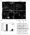
Figure 4. Activation of autophagy by ARF in U2-OS cells is p53-dependent
A, NARF cells transfected with p53 siRNA and induced with IPTG accumulate significantly fewer GFP-LC3 puncta compared with NARF cells transfected with control siRNA. Cells were transfected with siRNA, infected with adenovirus expressing GFP-LC3, induced with IPTG, and observed under a fluorescence microscope. B, quantitation of the percentage of cells with GFP-LC3 puncta after transfection with control or p53 siRNA. C, induction of p14ARF in NARF cells transfected with control siRNA causes a significant increase in p53 level and in the level of the p53 targets MDM2, p21, and PUMA, as detected by Western blot. The increase in the level of LC3-II is indicative of the activation of autophagy in these cells. In cells transfected with p53 siRNA, the level of p53 and of the p53 targets is undetectable, and there is no significant increase of LC3-II level.

References

    1. Sherr CJ. Divorcing ARF and p53: an unsettled case. Nat Rev Cancer. 2006;6:663–673. - PubMed
    1. Zhang Y, Xiong Y, Yarbrough WG. ARF promotes MDM2 degradation and stabilizes p53: ARF-INK4a locus deletion impairs both the Rb and p53 tumor suppression pathways. Cell. 1998;92:725–734. - PubMed
    1. Pomerantz J, Schreiber-Agus N, Liegeois NJ, et al. The Ink4a tumor suppressor gene product, p19Arf, interacts with MDM2 and neutralizes MDM2’s inhibition of p53. Cell. 1998;92:713–723. - PubMed
    1. Korgaonkar C, Zhao L, Modestou M, Quelle DE. ARF function does not require p53 stabilization or Mdm2 relocalization. Mol Cell Biol. 2002;22:196–206. - PMC - PubMed
    1. Chen D, Kon N, Li M, Zhang W, Qin J, Gu W. ARF-BP1/Mule is a critical mediator of the ARF tumor suppressor. Cell. 2005;121:1071–1083. - PubMed

Publication types

MeSH terms

Substances