. 2008 Feb;135(4):717-27.
doi: 10.1242/dev.015339. Epub 2008 Jan 16.

Inactivation of nuclear Wnt-beta-catenin signaling limits blastocyst competency for implantation

Affiliations

Inactivation of nuclear Wnt-beta-catenin signaling limits blastocyst competency for implantation

Huirong Xie et al. Development. 2008 Feb.

Abstract

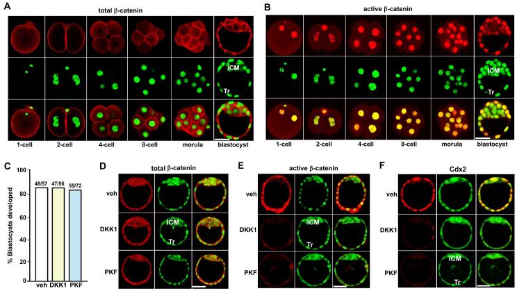
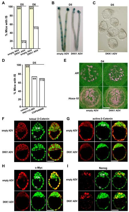
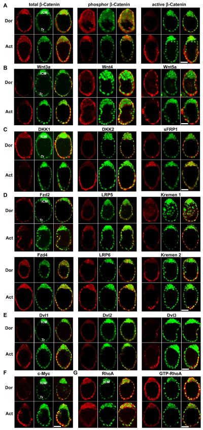
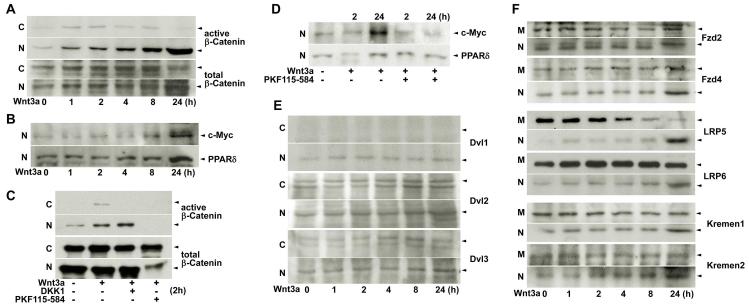
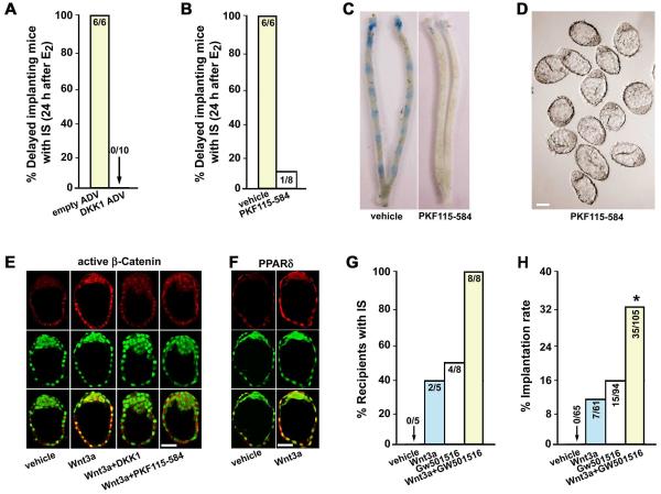
The activation of the blastocyst, a process by which it gains competency to attach with the receptive uterus, is a prerequisite for successful implantation. However, the molecular basis of blastocyst activation remains largely unexplored. Combining molecular, pharmacological and physiological approaches, we show here that silencing of Wnt-beta-catenin signaling in mice does not adversely affect the development of preimplantation embryos to blastocysts and uterine preparation for receptivity, but, remarkably, blocks blastocyst competency to implantation. Using the physiologically relevant delayed implantation model and trophoblast stem cells in culture, we further demonstrate that a coordinated activation of canonical Wnt-beta-catenin signaling with attenuation of the non-canonical Wnt-RhoA signaling pathway ensures blastocyst competency to implantation. These findings constitute novel evidence that Wnt signaling is at least one pathway that determines blastocyst competency for implantation.


Figures

Fig. 1
Fig. 1. Consequence of silencing nuclear β-catenin on mouse preimplantation embryo development
(A, B) Immunofluorescence localization of total and dephospho (active) β-catenin in mouse preimplantation embryos. (C-F) Recombinant DKK1 protein (5 μg/ml) and PKF115-584 (0.1 μM) block nuclear import of active dephospho β-catenin and Cdx2 expression in preimplantation embryos, without interfering with the cellular level of total β-catenin and the development of 2-cell embryos to blastocysts in culture. Two-cell embryos recovered by flushing day 2 pregnant oviducts were cultured in groups of 5-10 in 25 μl of M16 medium under silicon oil in an atmosphere of 5% CO2 and 95% air at 37□ for 72 h and the number of blastocysts developed was recorded. Experiments were repeated 3-5 times. The number above the bar in panel C indicates the number of blastocysts developed per the number of cultured 2-cell embryos. Images shown depict Cy3-labeled active β-catenin as red, SYTO-13-labeled nuclei as green and merge as yellow. Bar, 50 μm. ICM, inner cell mass; Tr, trophectoderm; veh: vehicle; PKF: PKF115-584.
Fig. 2
Fig. 2. Inactivation of Wnt-β-catenin signaling derails on-time implantation
(A) Implantation in mice receiving empty or DKK1 ADV on days 5 and 6 of pregnancy. Implantation sites (IS) were visualized by the blue dye method. Numbers within the bar indicate the number of mice with IS/total number of mice examined. (B, C) Representative photomicrograph of uteri with or without IS (blue bands) and recovered unimplanted morphologically normal blastocysts from those without blue reaction (Bar, 50 μm). (D) Implantation in mice receiving vehicle, PKF115-584 or CGP049090 (each 10 mg/Kg·BW) on day 5. Numbers within the bar indicate the number of mice with IS/total number of mice examined. (E) In situ hybridization showing comparable expressions of amphiregulin (AR) and Hoxa-10 in day 4 uteri of mice receiving empty or DKK1 ADV (Bar, 200 μm). (F-I) Overexpression of DKK1 via DKK1 ADV exerts no effects on the cellular level of total β-catenin, but remarkably attenuates nuclear stabilization of active dephospho β-catenin and c-Myc expression in blastocyst trophectoderms (Tr) when examined on day 4 midnight (day 4.5). In contrast, Nanog, an inner cell mass (ICM) marker gene, is normally expressed in ICM cells of blastocysts recovered from pregnant females receiving either DKK1 ADV or empty vectors on day 4.5. Represented immunofluorescence staining images depict Cy3-labeled antigens as red, SYTO-13-labeled nuclei as green and merge as yellow. Bar, 50 μm.
Fig. 3
Fig. 3. Wnt pathways in dormant and activated blastocysts
(A) Differential patterns of phospho (inactive) and dephospho (active) β-catenin during blastocyst activation. While total β-catenin distribution was comparable in dormant (Dor) and activated (Act) blastocysts, phospho β-catenin was primarily detected in the inner cell mass (ICM) and active β-catenin mostly in the trophectoderm (Tr) of activated blastocysts. (B) Wnt3a was induced in activated blastocyst Tr, while Wnt4 and Wnt5a were detected at high levels in the same cell-types in both dormant and activated blastocysts. (C) Dynamic expression of Wnt antagonists, DKK1, DKK2 and sFRP1 in delayed implanting blastocysts. While DKK1 was downregulated, DKK2 was induced in the Tr with blastocyst activation. In contrast, sFRP1 expression was restricted to the ICM of activated blastocysts. (D) Expression of Wnt receptor subtypes, Fzd 2, Fzd4, LRP5, LRP6, Kremen 1 and Kremen 2 in dormant and activated blastocysts. One intriguing observation is the internalization and nuclear import of Wnt receptors in activated blastocysts. (E) Expression of Dvl1-3 proteins in delayed implanting blastocysts. It was notable that Dvl1 and Dvl3 increasingly accumulated in the cytoplasm with visible nuclear localization of Dvl1 in the Tr during blastocyst activation. (F) c-Myc was induced in Tr cells of activated blastocysts. (G) Downregulation of total and GTP-bounded (active) RhoA GTPase in the Tr during blastocyst activation. Represented immunofluorescence staining images shown depict Cy3-labeled antigens as red, SYTO-13-labeled nuclei as green and merge as yellow. Bar, 50 μm.
Fig. 4
Fig. 4. Nuclear β-catenin signaling in TS cells in culture
(A) Time-dependent accumulation and nuclear translocation of active β-catenin in response to recombinant Wnt3a protein (50 ng/ml) in differentiating TS cells. (B) Wnt3a induced c-Myc and PPARδ expression in differentiating TS cells. (C) DKK1 (1 μg/ml) and PKF115-584 (1 μM) blocked Wnt3a-induced cytoplasmic accumulation of active dephospho β-catenin in TS cells by 2 hour of cotreatments. Notably, even basal levels of nuclear β-catenin disappeared following PKF115-584 treatment. (D) Similar treatments with DKK1 (1 μg/ml) and PKF115-584 (1μM) downregulated Wnt3a-induced c-Myc and PPARδ expression in differentiating TS cells. (E) Dvl1-3 proteins in TS cells. While Dvl1 was only detected in the nucleus, Dvl2 and Dvl 3 were detected in both the cytoplasm and nucleus in response to Wnt3a. (F) Wnt receptors, Fzd 2, Fzd4, LRP5, LRP6, Kremen 1 and Kremen 2 in TS cells. Wnt3a facilitated nuclear import of Wnt receptor subtypes in differentiating TS cells, mimicking the finding in blastocysts during activation. Representative pictures of Western blotting analysis of Wnt family components in TS cells were presented in panels A-F. C, M or N, cytoplasmic, membrane or nuclear protein extraction.
Fig. 5
Fig. 5. Wnt-β-catenin signaling synergizes with that of PPARδ to confer blastocyst competency for implantation
(A, B) Overexpressing levels of DKK1 or PKF115-584 blocked activation of dormant blastocysts for implantation in response to E2 (3 ng/mouse). Numbers within the bar indicate the number of mice with implantation sites (IS)/total number of mice examined. (C, D) Representative photomicrographs of uteri without blue bands and morphologically dormant blastocysts recovered from mice treated with PKF115-584 (Bar, 50 μm). (E, F) Recombinant Wnt3a protein (200 ng/ml) induced nuclear stabilization of active dephospho β-catenin and PPARδ expression in dormant blastocysts in culture. Cotreatment of Wnt3a with DKK1 (1 μg/ml) or PKF115-584 (1 μM) antagonized Wnt3a-induced β-catenin stabilization. Images shown depict Cy3-labeled antigens as red, SYTO-13-labeled nuclei as green and merge as yellow. Bar, 50 μm. (G, H) Wnt3a and/or GW501516 conferred blastocyst implantation competency. Dormant blastocysts were cultured in the presence of vehicle, Wnt3a (200 ng/ml) and/or GW501516 (a selective PPARδ agonist, 1 μM) for 24h before transferred into pseudopregnant delayed recipients. Numbers within the bar in G indicate the number of recipients with IS/total number of mice examined, and those in H indicate the number of IS/total number of blastocysts transferred.

References

    1. Bafico A, Liu G, Yaniv A, Gazit A, Aaronson SA. Novel mechanism of Wnt signalling inhibition mediated by Dickkopf-1 interaction with LRP6/Arrow. Nat Cell Biol. 2001;3:683–6. - PubMed
    1. Barrow JR. Wnt/PCP signaling: a veritable polar star in establishing patterns of polarity in embryonic tissues. Semin Cell Dev Biol. 2006;17:185–93. - PubMed
    1. Berdeaux RL, Diaz B, Kim L, Martin GS. Active Rho is localized to podosomes induced by oncogenic Src and is required for their assembly and function. J Cell Biol. 2004;166:317–23. - PMC - PubMed
    1. Braga VM, Machesky LM, Hall A, Hotchin NA. The small GTPases Rho and Rac are required for the establishment of cadherin-dependent cell-cell contacts. J Cell Biol. 1997;137:1421–31. - PMC - PubMed
    1. Capelluto DG, Kutateladze TG, Habas R, Finkielstein CV, He X, Overduin M. The DIX domain targets dishevelled to actin stress fibres and vesicular membranes. Nature. 2002;419:726–9. - PubMed

Publication types

MeSH terms