. 2008 Mar;82(6):2938-51.
doi: 10.1128/JVI.02126-07. Epub 2008 Jan 16.

Targets of small interfering RNA restriction during human immunodeficiency virus type 1 replication

Affiliations

Targets of small interfering RNA restriction during human immunodeficiency virus type 1 replication

Yong Gao et al. J Virol. 2008 Mar.

Abstract

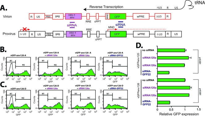
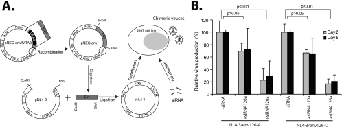
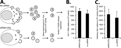
Small interfering RNAs (siRNAs) have been shown to effectively inhibit human immunodeficiency virus type 1 (HIV-1) replication in vitro. The mechanism(s) for this inhibition is poorly understood, as siRNAs may interact with multiple HIV-1 RNA species during different steps of the retroviral life cycle. To define susceptible HIV-1 RNA species, siRNAs were first designed to specifically inhibit two divergent primary HIV-1 isolates via env and gag gene targets. A self-inactivating lentiviral vector harboring these target sequences confirmed that siRNA cannot degrade incoming genomic RNA. Disruption of the incoming core structure by rhesus macaque TRIM5alpha did, however, provide siRNA-RNA-induced silencing complex access to HIV-1 genomic RNA and promoted degradation. In the absence of accelerated core disruption, only newly transcribed HIV-1 mRNA in the cytoplasm is sensitive to siRNA degradation. Inhibitors of HIV-1 mRNA nuclear export, such as leptomycin B and camptothecin, blocked siRNA restriction. All HIV-1 RNA regions and transcripts found 5' of the target sequence, including multiply spliced HIV-1 RNA, were degraded by unidirectional 3'-to-5' siRNA amplification and spreading. In contrast, HIV-1 RNA 3' of the target sequence was not susceptible to siRNA. Even in the presence of siRNA, full-length HIV-1 RNA is still encapsidated into newly assembled viruses. These findings suggest that siRNA can target only a relatively "naked" cytoplasmic HIV-1 RNA despite the involvement of viral RNA at nearly every step in the retroviral life cycle. Protection of HIV-1 RNA within the core following virus entry, during encapsidation/virus assembly, or within the nucleus may reflect virus evolution in response to siRNA, TRIM5alpha, or other host restriction factors.


Figures

FIG. 1.
FIG. 1.
Mapping the potentially siRNA degradation-susceptible HIV-1 RNA species during the retroviral life cycle. As illustrated in panel A, siRNA complexed to RISC may interfere with several stages of the HIV-1 life cycle to inhibit virus replication: siRNA may inhibit following virus entry by targeting HIV-1 genomic RNA (1); siRNA may inhibit transcription by directing chromatin modification (2); siRNA may degrade viral mRNA in the nucleus (3); siRNA may degrade viral mRNA following export from the nucleus to the cytoplasm (4); siRNA (through an miRNA form) may reduce translation of viral proteins (5); siRNA may interfere with the packaging of viral genomic RNA into a new virus particle or siRNA-RISC could be incorporated into the budding virus (6). PIC, pre-integration complex. Panel B delineates the sequence and target sites of the seven siRNAs designed to specifically inhibit HIV-1 v120-A (a subtype A virus) or v126-D (a subtype D virus). The locations and sequence complementarity of each siRNA in the HIV-1 env or gag genes are depicted in panel B. The starting nucleotide position (HXB2 numbering) is provided for each siRNA.
FIG. 2.
FIG. 2.
Efficiency and specificity of siRNA-mediated inhibition of divergent HIV-1 isolates. Panel A provides the drug susceptibility curves after 5 days of v120-A (V120-UG, where UG is Uganda) replication in U87.CD4.CXCR4 cells in the presence of various concentrations (0.0002 to 20 nM) of siRNA120a, siRNA120b, siRNA120c, and siRNA120d (transfected by Lipofectamine 2000). (B) v126-D replication in U87.CD4.CXCR4 cells treated with siRNA126a and siRNA126b. (C) The specificity of siRNA120a was measured in U87.CD4.CXCR4 cells exposed to v120-A and v126-D (V126-UG). (D) Finally, the decay of antiviral activity of siRNA120a in U87.CD4.CXCR4 cells exposed to v120-A was determined over a 5- to 8-day period. Each infection and siRNA treatment were performed in triplicate; in order to improve the clarity of panels A and B, error bars of ∼10% variance are not shown.
FIG. 3.
FIG. 3.
Effect of specific siRNAs on viral RNA and cDNA synthesis. (A) v120-A RNA and proviral DNA were measured by env-specific RT-PCR and PCR (respectively) in siRNA-treated U87.CD4.CXCR4 cells at various time points after infection and treatment with siRNAcontrol (siRNAcon), 3TC, siRNA120a, and siRNAgag1. PCR amplifications of undiluted and diluted nucleic acid extracts from infected cells are shown on the left of panel A, and quantitation of these PCR products is shown on the right. The production of HIV-1 DNA following these infections was also measured by real-time PCR specific for the 5′ LTR region (panel B). Panel C provides the relative production of v120-A under these various conditions as measured by RT activity released in the supernatant at different time points postinfection. All experiments were performed in triplicate. Since variance was less than 10% for relative virus production and siRNA inhibition, RT-PCR amplifications were performed only on a set of serially diluted RNA samples.
FIG. 4.
FIG. 4.
Determining the sensitivity of incoming genomic HIV-1 RNA to siRNA degradation. (A) The env gene of v120-A or v126-D was inserted into the self-inactivating lentiviral vector pMND-GFPpre, which contains an internal MND promoter driving GFP, the central polypurine tract (cPPT)/central termination sequence, and the HIV-1 LTR with U3 deleted and cytomegalovirus promoter added at the 5′ LTR. PBS, primer binding sequence; wPRE, Woodchuck hepatitis virus posttranscriptional regulatory element. The virus particles derived from triple transfections of 293T cells with the packaging vector (pCMVΔR8.91), the VSV-G-pseudotyping vector (pMD.G), and pMND.GFPpre, pGFPenv120-A (containing the env target sequence of v120-D), or pGFPenv126-D (containing the env target sequence of v126-D). The viral RNA in these virus particles can be reverse transcribed, and the proviral DNA can be integrated into the target 293T cells. However, the LTR in the integrated virus genome cannot drive transcription and thus does not express viral RNA with the env target sequence. An internal promoter in the integrated virus genome can transcribe an mRNA encoding enhanced GFP. Eight graphs describe the flow cytometry results gated for 293T cells expressing GFP (M2) from the vGFP-env120-A (B) and vGFP-env126-D (C) infections in the presence or absence of siRNA120a or siRNA126a. Quantitative results from this experiment are shown in panel D. All experiments were performed in triplicate.
FIG. 5.
FIG. 5.
siRNA degradation of genomic HIV-1 RNA following disruption of the virus cores by TRIM5αrh. lipo, Lipofectamine 2000. (A) VSV envelope-pseudotyped virus vGFP-env120-A was used to infect 293T or 293TTRIM5αrh cells in the presence or absence of 20 nM siRNA120a or siRNA126a. GFP-expressing cells were measured by flow cytometry. Semiquantitative RT-PCR was performed on extracted RNA from the cells exposed to vGFP-env120-A with or without siRNAs. Panels B and C show the quantified results from the experiments using the 293T or 293TTRIM5αrh cells, performed in triplicate. Panel D displays the relative amounts of HIV env mRNA that were RT-PCR amplified at 8 h and 24 h or of HIV-1 env DNA that were PCR amplified at 36 h and 48 h after virus addition.
FIG. 6.
FIG. 6.
siRNA inhibition of HIV-1 transcription for cells transfected with proviral DNA vectors. Panel A provides a schematic representation of the general cloning strategy to replace the env gene in pNL4-3 with the env genes of v120-A and v126-D. The resulting plasmids were then transfected in 293T cells in the presence or absence of siRNAs. Amp, ampicillin resistance cassette; PCMV, cytomegalovirus promoter; Zeo, Zeocin resistance cassette. (B) The relative virus production as measured by RT activity was monitored in supernatants at days 2 and 5 after transfection and siRNA treatment.
FIG. 7.
FIG. 7.
siRNA degradation on different HIV-1 mRNA species in the presence or absence of inhibitors of nuclear mRNA export. Panel A provides a schematic illustration of the location of the RT-PCR primers in the HIV-1 genome and in specific HIV-1 unspliced and multiply spliced mRNA species. Images of the agarose gels containing the various amplified HIV-1 RNA products are shown in panel B. RNA for these RT-PCRs was extracted from pNL4-3/env120-A-transfected 293T cells exposed to siRNA120a in the presence or absence of inhibitors of the CRM-1 nuclear export pathway (leptomycin B [LepB] and camptothecin [Camp]). The relative levels of these various HIV-1 RNA messages (measured by RT-PCR as described for panels A and B) in the absence of siRNAs (set to 100%) were compared to levels in the presence of siRNA and with no inhibitor of nuclear export (C), leptomycin B (D), or camptothecin (E). All experiments whose results are shown in panels A through E were performed in triplicate. Since variance was less than 10% for relative virus production and siRNA inhibition, RT-PCR amplifications were performed only on a set of serially diluted RNA samples. Panel F provides a comparison of HIV-1 RNA levels determined by using semiquantitative PCR and real-time PCR methods (SYBR green). (G) The levels of HIV-1 RNA were determined by real-time PCR for each replicate in this triplicate experiment. The levels of HIV-1 RNA were measured in the absence of nuclear export inhibitor, with leptomycin B, or with camptothecin by using the RNA protection method described in Materials and Methods. Two probes specific for HIV-1 RU5 (unspliced RNA) or U3R products were used in this RPA. semi-quant, semiquantitative.
FIG. 8.
FIG. 8.
Measuring HIV-1 protein production in cells and supernatant in the presence of siRNAs. CA p24 or unprocessed p55gag was monitored by Western blotting for cells (C) and supernatants (A and B), at days 2 and 5, of 293T cells transfected with pNL4-3/env120-A or pNL4-3/env126-D and treated with siRNA120a or siRNA126a or mock treated. Both panels A and C employed a 1:400 dilution of patient sera derived from individuals infected with HIV-1 of either subtype A (JLT02A) or subtype C (JCR12) as a primary anti-p24 antibody, followed by peroxidase-conjugated secondary antibodies. (C) The anti-gp120 B13 antibody was employed in Western blot analyses with virus lysates obtained from day 5 supernatants of 293T cells transfected with pNL4-3/env and mock treated or treated with siRNA120a or siRNA126a. The B13 anti-gp120 antibody binds to a highly conserved linear epitope found in most HIV-1 gp120 sequences of most subtypes (panel B) (1)
FIG. 9.
FIG. 9.
siRNA effects on packaging of genomic HIV-1 RNA and on the infectivity of newly produced virus. Panel A provides a schematic of virus production in U87.CD4.CXCR4 cells in the presence and absence of siRNAs. Although siRNAs inhibit 80 to 90% of virus production, the v120-A released in the supernatant in the presence or absence of siRNA120a was serially diluted and quantified by RT activity to determine a “virtual” TCID50 (36). (B) Equalized infectious titers (based on RT activity) of virus (with or without siRNA) were again serially diluted and used to infect U87.CD4.CXCR4 cells in the absence of siRNA to determine if siRNA have any inhibitory effects on infectivity of newly produced virus. The HIV-1 RNA levels in virus particles equalized for RT activity following production from cells infected in the presence or absence of siRNA. HIV-1 RNA levels were measured with real-time PCR using an ABI Prism 7000 sequence detection system. All conditions were performed in triplicate.

Similar articles

Cited by

References

    1. Abacioglu, Y. H., T. R. Fouts, J. D. Laman, E. Claassen, S. H. Pincus, J. P. Moore, C. A. Roby, R. Kamin-Lewis, and G. K. Lewis. 1994. Epitope mapping and topology of baculovirus-expressed HIV-1 gp160 determined with a panel of murine monoclonal antibodies. AIDS Res. Hum. Retrovir. 10371-381. - PubMed
    1. Abraha, A., R. M. Troyer, M. E. Quinones-Mateu, and E. J. Arts. 2005. Methods to determine HIV-1 ex vivo fitness. Methods Mol. Biol. 304355-368. - PubMed
    1. Arhel, N. J., S. Souquere-Besse, S. Munier, P. Souque, S. Guadagnini, S. Rutherford, M. C. Prevost, T. D. Allen, and P. Charneau. 2007. HIV-1 DNA Flap formation promotes uncoating of the pre-integration complex at the nuclear pore. EMBO J. 263025-3037. - PMC - PubMed
    1. Arts, E. J., X. Li, Z. Gu, L. Kleiman, M. A. Parniak, and M. A. Wainberg. 1994. Comparison of deoxyoligonucleotide and tRNA(Lys-3) as primers in an endogenous human immunodeficiency virus-1 in vitro reverse transcription/template-switching reaction. J. Biol. Chem. 26914672-14680. - PubMed
    1. Arts, E. J., J. Mak, L. Kleiman, and M. A. Wainberg. 1994. DNA found in human immunodeficiency virus type 1 particles may not be required for infectivity. J. Gen. Virol. 751605-1613. - PubMed

Publication types