Review
. 2008 Feb;29(1):62-75.
doi: 10.1210/er.2007-0004. Epub 2008 Jan 16.

The role of membrane glycoprotein plasma cell antigen 1/ectonucleotide pyrophosphatase phosphodiesterase 1 in the pathogenesis of insulin resistance and related abnormalities

Affiliations
Review

The role of membrane glycoprotein plasma cell antigen 1/ectonucleotide pyrophosphatase phosphodiesterase 1 in the pathogenesis of insulin resistance and related abnormalities

Ira D Goldfine et al. Endocr Rev. 2008 Feb.

Erratum in

  • Endocr Rev. 2009 Feb;30(1):117

Abstract

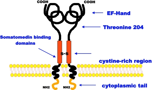
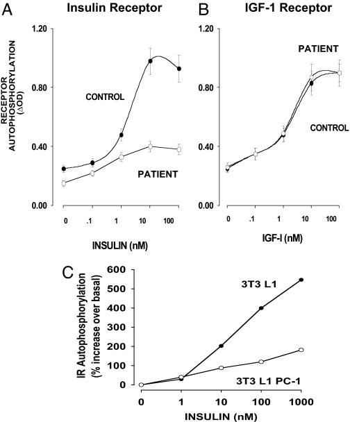
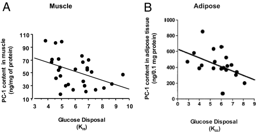
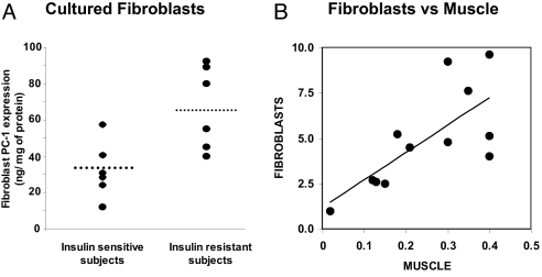
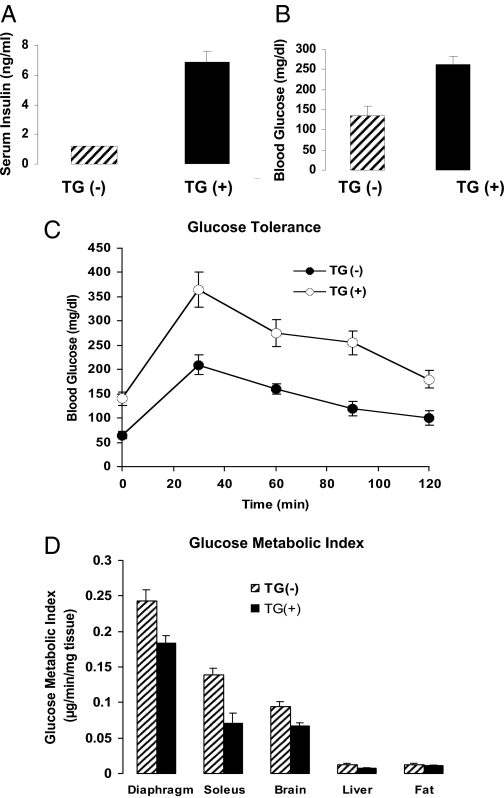
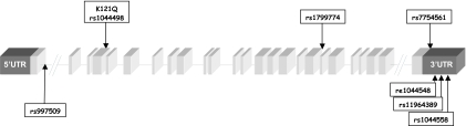
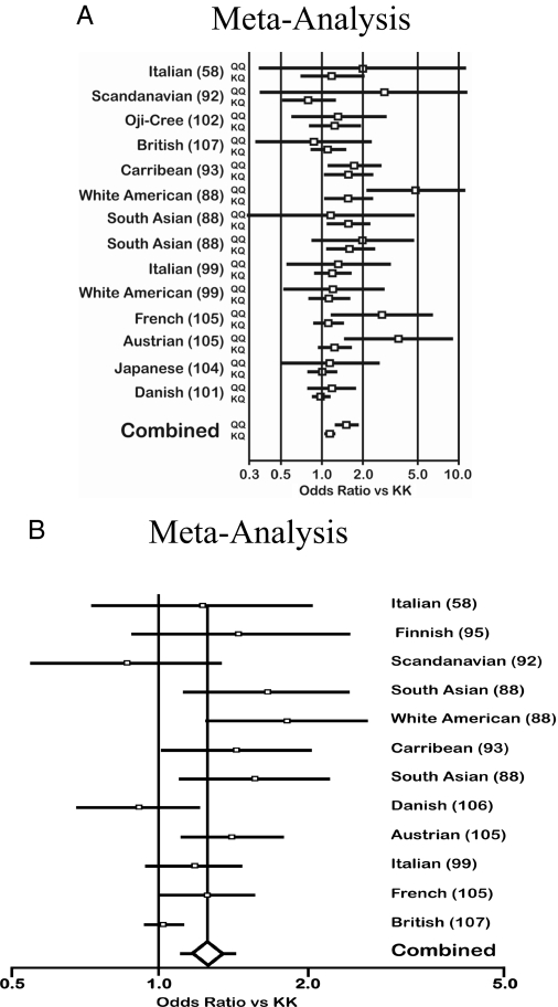
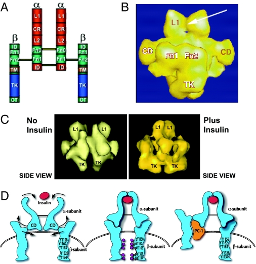
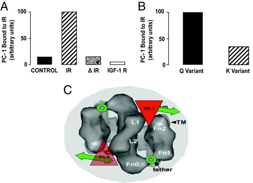
Insulin resistance is a major feature of most patients with type 2 diabetes mellitus (T2D). A number of laboratories have observed that PC-1 (membrane [corrected] glycoprotein plasma cell antigen 1; also termed [corrected] ectonucleotide pyrophosphatase phosphodiesterase 1 or ENPP1) [corrected] is either overexpressed or overactive in muscle, adipose tissue, fibroblasts, and other tissues of insulin-resistant individuals, both nondiabetic and diabetic. Moreover, PC-1 (ENPP1) overexpression [corrected] in cultured cells in vitro and in transgenic mice in vivo, [corrected] impairs insulin stimulation of insulin receptor (IR) activation and downstream signaling. PC-1 binds to the connecting domain of the IR alpha-subunit that is located in residues 485-599. The connecting domain transmits insulin binding in the alpha-subunit to activation of tyrosine kinase activation in the beta-subunit. When PC-1 is overexpressed, it inhibits insulin [corrected]induced IR beta-subunit tyrosine kinase activity. In addition, a polymorphism of PC-1 (K121Q) in various ethnic populations is closely associated with insulin resistance, T2D, and cardio [corrected] and nephrovascular diseases. The product of this polymorphism has a 2- to 3-fold increased binding affinity for the IR and is more potent than the wild-type PC-1 protein (K121K) in inhibiting the IR. These data suggest therefore that PC-1 is a candidate protein that may play a role in human insulin resistance and T2D by its overexpression, its overactivity, or both.


Figures

Figure 1
Figure 1
Structure of PC-1. Major domains of the molecule are shown. EF-hand, Ca+2 binding domain that maintains structure; Threonine 204, phosphodiesterase/pyrophosphatase site. The somatomedin-like domains are located near the plasma membrane and appear to be involved in interacting with the IR. The high cysteine region is involved in dimer formation.
Figure 2
Figure 2
PC-1 inhibition of IR β-subunit autophosphorylation in fibroblasts. A and B, Skin fibroblasts from the original patient with a 4-fold elevation of PC-1 were stimulated with either insulin (A) or IGF-I (B) for 5 min, and IR and IGF-IR autophosphorylation was measured by specific ELISAs. [Derived from Ref. .] C, 3T3 L1 mouse fibroblasts were transfected to overexpress human PC-1 4-fold. IR autophosphorylation in control 3T3 L1 and 3T3 L1 PC-1 fibroblasts was measured by specific IR ELISA.
Figure 3
Figure 3
Correlation between muscle and adipose PC-1 levels with in vivo insulin action. A, Correlation between PC-1 content in skeletal muscle of 28 nonobese, nondiabetic subjects and in vivo insulin sensitivity measured by the iv insulin tolerance test Kitt (51) (r = 0.51, P < 0.035) and positively correlated with plasma insulin levels. B, Correlation between PC-1 content in human adipose tissue of 19 nonobese, nondiabetic subjects and in vivo insulin sensitivity as indicated by Kitt values. PC-1 content negatively correlated with insulin sensitivity (r = −0.57, P = 0.011) and positively correlated with plasma insulin levels. [Panel A adapted from L. Frittitta et al.: Diabetologia 39:1190–1195, 1996 (51) with kind permission from Springer Science and Business Media. Panel B adapted from L. Frittitta et al.: Diabetologia 40:282–289, 1997 (62) with kind permission from Springer Science and Business Media.]
Figure 4
Figure 4
Fibroblast PC-1 levels. A, PC-1 content in cultured human skin fibroblasts from “normal” (healthy nonobese, nondiabetic) subjects with different insulin sensitivity (n = 12). Primary cultures of fibroblasts were established from 4-mm forearm skin biopsies. Horizontal bars indicate mean values. P = 0.01, sensitive vs. resistant subjects. B, Correlation between PC-1 content in cultured human skin fibroblasts from “normal” (healthy nonobese, nondiabetic) subjects and PC-1 content in skeletal muscle obtained from the same subjects. Over a wide range of PC-1 values, the content of PC-1 in skin fibroblasts closely reflected its content in muscle tissue (P = 0.01). [Panel A adapted from Frittitta et al.:Diabetes 47:1096–1100, 1998 (63) with permission from The American Diabetes Association. Panels A and B adapted from Goldfine et al.: Ann NY Acad Sci 892:204–222, 1999 (56).]
Figure 5
Figure 5
Studies in PC-1 transgenic mice. Insulin (A) and glucose (B) levels in 10 PC-1 transgenic (TG+) vs. 10 control mice (TG−). C, Glucose tolerance tests (five per group). D, Glucose metabolic index in tissues of transgenic animals overexpressing PC-1. Animals were given a constant insulin infusion, and the uptake of 2-[14C]DG was measured in various tissues (seven control and five transgenic). In transgenic animals, glucose uptake (Rg) was significantly decreased in diaphragm and soleus muscles and brain. [Adapted with permission from B. A. Maddux et al.: Am J Physiol Endocrinol Metab 290:E746–E749, 2006 (57).]
Figure 6
Figure 6
Genomic structure PC-1. The 5′ and 3′ UTRs are shown in dark gray. The 25 exons of the PC-1 gene are shown in light gray. The several single nucleotide polymorphisms so far reported to be associated with insulin resistance and related abnormalities are indicated.
Figure 7
Figure 7
Meta-analyses of the K121Q polymorphism. Meta-analyses on the association between the PC-1 K121Q polymorphism and T2D suggesting a significant diabetogenic role of this in modulating the risk of diabetes. Meta-analysis A, Ref. ; meta-analysis B, Ref. . For both, the odds ratios vs. K121K allele are calculated with a 95% confidence interval. In A, the KQ and QQ genes are shown separately. In both A and B, a significantly increased risk is given by each K121Q polymorphism. [Meta-analysis A adapted from N. Grarup et al.: Diabetologia 49:2097–2104, 2006 ( with kind permission of Springer Science and Business Media; B adapted from M. N. Weedon et al.: Diabetes 55:3175–3179, 2006 ( with permission from The American Diabetes Association.]
Figure 8
Figure 8
Traditional and STEM models of the IR. A, Traditional model of the IR with the major domains shown. The following regions are indicated: α-subunit domains: L1, long 1; CR, cysteine-rich; L2, long 2; α Fn 0, α fibronectin 0; α Fn 1, α Fibronectin 1; ID, insert-domain. β-subunit domains: ID, insert-domain; β Fn 1, β fibronectin 1; β Fn 2, β fibronectin 2; TM, transmembrane; TK, tyrosine-kinase; CT, C-terminal. B, Three-dimensional reconstruction (side view) from STEM. The arrow indicates the insulin-binding site at the L1 domains. The Fn 1, Fn 2, and TK domains of the β-subunit are shown. C, STEM data showing the effect of insulin binding to move the β-subunits. D, Model of proposed activation of the IR β-subunit by the CD. The CD is a connecting domain in the IR α-subunit that includes residues 485–599. In this model insulin binds to the α-subunit to activate the CDs which then move the β-subunits together to facilitate transphosphorylation. When PC-1 binds to the CD it blocks the movement of the β-subunits induced by insulin. [Derived from C. C. Yip and P. Ottensmeyer: Biol Chem 278:27329–27332, 2003 (126).]
Figure 9
Figure 9
PC-1 associates with the IR and blocks IR function: STEM model of PC-1 inhibition of the IR. A. Four types of cultured HTC cells transfected with the K121 of PC-1 were employed. [Derived from Ref. .] Control, Cells with low numbers of endogenous IRs; IR, cells transfected with large numbers of normal human IRs; Δ 485–599, cells transfected with large numbers of human IRs having a deletion of the IR at residues 485–599 in the α-subunit; and IGF-IR, cells transfected with large numbers of the human IGF-IR. PC-1 bound to either the IR or the IGF-IR was measured by ELISA. B, Comparison of binding of the K121 and Q121 allele of PC-1 to the IR. HTC cells, transfected with equal amounts of either the K or Q variants of PC-1, were also cotransfected with the IR. PC-1 bound to the IR was measured by ELISA. C, STEM data fitting PC-1 into the IR model to visualize its putative inhibition of IR function. Extracellular top view of the IR dimer is presented showing the calculated location of PC-1 binding to the Fn0 region interacting with the Fn2 regions. In this location, PC-1 would act to inhibit the Fn1/Fn2 domains from interacting with the associated intracellular TK domains and thus prevent them from rotating toward each other to carry out TK transphosphorylation (covered portion of double-headed arrows). The “hinge” region between Fn0 and Fn1 is indicated by a circle and labeled “tether”.

Similar articles

Cited by

References

    1. Yeni-Komshian H, Carantoni M, Abbasi F, Reaven GM 2000 Relationship between several surrogate estimates of insulin resistance and quantification of insulin-mediated glucose disposal in 490 healthy nondiabetic volunteers. Diabetes Care 23:171–175 - PubMed
    1. Bogardus C, Lillioja S, Mott DM, Hollenbeck C, Reaven G 1985 Relationship between degree of obesity and in vivo insulin action in man. Am J Physiol 248:E286–E291 - PubMed
    1. Lillioja S, Mott DM, Zawadzki JK, Young AA, Abbott WG, Knowler WC, Bennett PH, Moll P, Bogardus C 1987 In vivo insulin action is familial characteristic in nondiabetic Pima Indians. Diabetes 36:1329–1335 - PubMed
    1. Warram JH, Martin BC, Krolewski AS, Soeldner JS, Kahn CR 1990 Slow glucose removal rate and hyperinsulinemia precede the development of type II diabetes in the offspring of diabetic parents. Ann Intern Med 113:909–915 - PubMed
    1. Lillioja S, Mott DM, Spraul M, Ferraro R, Foley JE, Ravussin E, Knowler WC, Bennett PH, Bogardus C 1993 Insulin resistance and insulin secretory dysfunction as precursors of non-insulin-dependent diabetes mellitus. Prospective studies of Pima Indians. N Engl J Med 329:1988–1992 - PubMed

Publication types

MeSH terms

Substances