. 2008 Mar 28;283(13):8496-507.
doi: 10.1074/jbc.M710433200. Epub 2008 Jan 16.

A proton-mediated conformational shift identifies a mobile pore-lining cysteine residue (Cys-561) in human concentrative nucleoside transporter 3

Affiliations

A proton-mediated conformational shift identifies a mobile pore-lining cysteine residue (Cys-561) in human concentrative nucleoside transporter 3

Melissa D Slugoski et al. J Biol Chem. .

Abstract

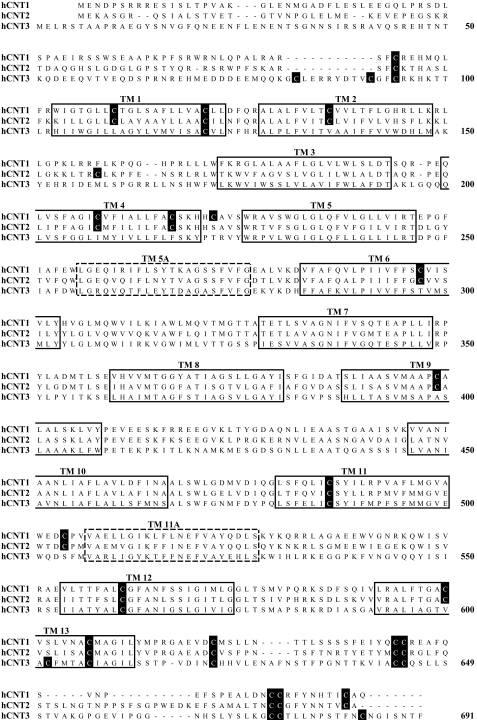
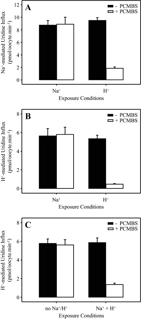
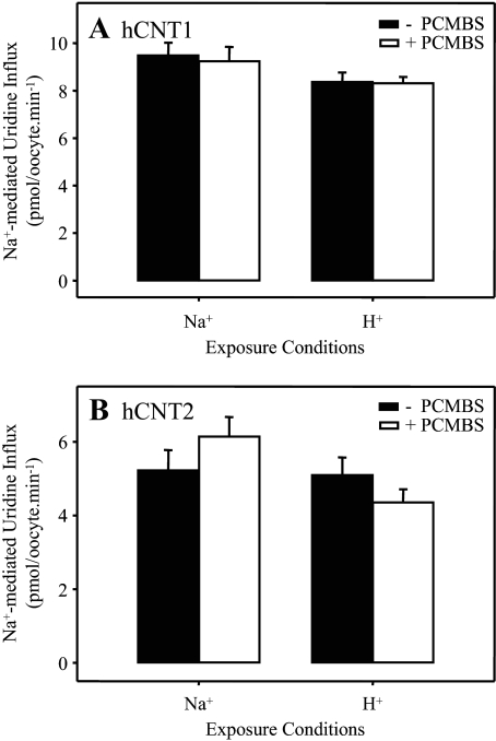
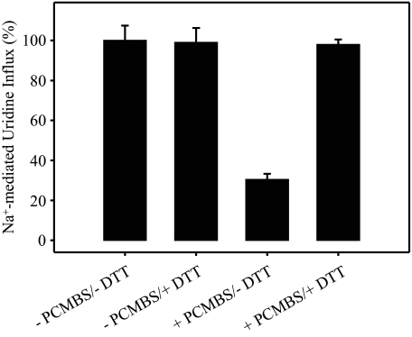
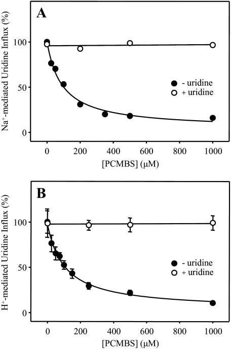
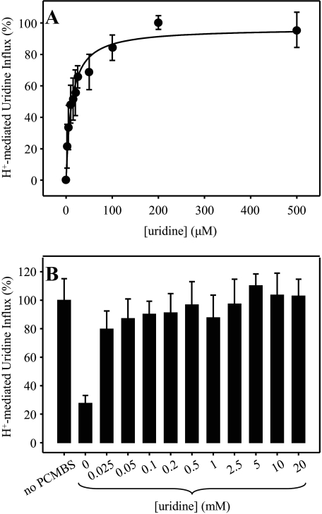
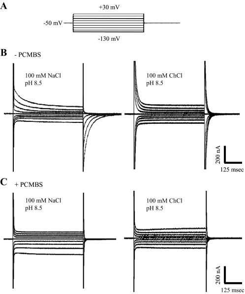
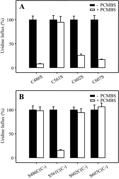
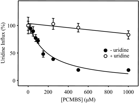
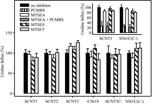
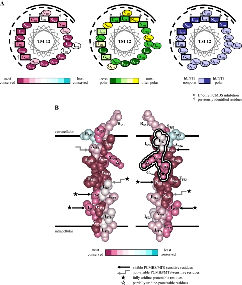
The concentrative nucleoside transporter (CNT) protein family in humans is represented by three members, hCNT1, hCNT2, and hCNT3. Belonging to a CNT subfamily phylogenetically distinct from hCNT1/2, hCNT3 mediates transport of a broad range of purine and pyrimidine nucleosides and nucleoside drugs, whereas hCNT1 and hCNT2 are pyrimidine and purine nucleoside-selective, respectively. All three hCNTs are Na(+)-coupled. Unlike hCNT1/2, however, hCNT3 is also capable of H(+)-mediated nucleoside cotransport. Using site-directed mutagenesis in combination with heterologous expression in Xenopus oocytes, we have identified a C-terminal intramembranous cysteine residue of hCNT3 (Cys-561) that reversibly binds the hydrophilic thiol-reactive reagent p-chloromercuribenzene sulfonate (PCMBS). Access of this membrane-impermeant probe to Cys-561, as determined by inhibition of hCNT3 transport activity, required H(+), but not Na(+), and was blocked by extracellular uridine. Although this cysteine residue is also present in hCNT1 and hCNT2, neither transporter was affected by PCMBS. We conclude that Cys-561 is located in the translocation pore in a mobile region within or closely adjacent to the nucleoside binding pocket and that access of PCMBS to this residue reports a specific H(+)-induced conformational state of the protein.


Figures

FIGURE 1.
FIGURE 1.
Cysteine residues in hCNT1, hCNT2, and hCNT3. An alignment of the amino acid sequences of hCNT1, hCNT2, and hCNT3 (GenBank™ accession numbers AAB53839, AAB88539, and AAG22551, respectively). The positions of 13 putative TMs are indicated by solid boxes. Two additional TMs present in an alternative 15 TM model of hCNT membrane architecture are indicated by dashed boxes. Cysteine residues are shown on black squares. Numbers refer to hCNT3 residue positions.
FIGURE 2.
FIGURE 2.
PCMBS inhibition of hCNT3. hCNT3-mediated influx of 20 μm [14C]uridine in Na+-containing (A) or H+-containing (B and C) medium (100 mm NaCl, pH 8.5, or 100 mm ChCl, pH 5.5, respectively; 1 min at 20 °C) was measured after 10 min of incubation on ice in the absence (solid bars) or presence (open bars) of 500 μm PCMBS in media containing Na+ but not H+ (100 mm NaCl, pH 8.5), H+ but not Na+ (100 mm ChCl, pH 5.5), lacking both Na+ and H+ (100 mm ChCl, pH 8.5), or containing both Na+ and H+ (100 mm NaCl, pH 5.5) as indicated. Values are corrected for basal non-mediated uptake in control water-injected oocytes and are the means ± S.E. of 10–12 oocytes.
FIGURE 3.
FIGURE 3.
PCMBS insensitivity of hCNT1 and hCNT2. Influx of 20 μm [14C]uridine in the presence of Na+ (100 mm NaCl, pH 7.5; 1 min at 20 °C) was measured in oocytes producing hCNT1 (A) or hCNT2 (B) after 10 min of incubation on ice in the absence (solid bars) or presence (open bars) of 500 μm PCMBS in media containing either Na+ but not H+ (100 mm NaCl, pH 8.5) or H+ but not Na+ (100 mm ChCl, pH 5.5) as indicated. Values are corrected for basal non-mediated uptake in control water-injected oocytes and are the means ± S.E. of 10–12 oocytes.
FIGURE 4.
FIGURE 4.
Reversal of PCMBS inhibition of hCNT3-mediated uridine uptake by DTT. hCNT3-expressing oocytes were incubated in the absence or presence of 500 μm PCMBS (100 mm ChCl, pH 5.5; 10 min on ice) followed by a second incubation in the absence or presence of 5 mm DTT (100 mm ChCl, pH 7.5; 1 min at 20 °C) before measuring uptake of 20 μm [14C]uridine in Na+-containing transport medium (100 mm NaCl, pH 7.5; 1 min at 20 °C). Data are presented as mediated transport, calculated as uptake in RNA-injected oocytes minus uptake in water-injected oocytes and are normalized to the respective influx of uridine in the absence of PCMBS and DTT (12.8 ± 1.0 pmol/oocyte·min-1). Each value is the mean ± S.E. of 10–12 oocytes.
FIGURE 5.
FIGURE 5.
PCMBS inhibition of hCNT3: concentration dependence and uridine protection. Influx of 20 μm [14C]uridine in both Na+- and H+-containing media (A and B, respectively) was measured after hCNT3-producing oocytes were incubated with various concentrations of PCMBS under acidic conditions either in the absence (solid circles) or in the presence (open circles) of 20 mm extracellular uridine as described in Fig. 2. Data are presented as mediated transport, calculated as uptake in RNA-injected oocytes minus uptake in water-injected oocytes, and are normalized to the respective influx of uridine in the absence of inhibitor (8.7 ± 0.5 (A) and 5.3 ± 0.7 (B) pmol/oocyte·min-1). Each value is the mean ± S.E. of 10–12 oocytes. Error bars are not shown where values were smaller than that represented by the symbols.
FIGURE 6.
FIGURE 6.
PCMBS inhibition of hCNT3: concentration dependence of uridine protection. Influx of 20 μm [3H]uridine in H+-containing medium was measured in oocytes producing hCNT3 after incubation with 500 μm PCMBS under acidic conditions in the presence of 0–500 μm (A) or 0–20 mm (B) extracellular uridine as described in Fig. 2. Data are presented as mediated transport, calculated as uptake in RNA-injected oocytes minus uptake in water-injected oocytes, and normalized to the respective influx of uridine in the absence of inhibitor (8.3 ± 1.3 (A) and 7.3 ± 0.6 (B) pmol/oocyte·min-1). Each value is the mean ± S.E. of 10–12 oocytes. Error bars are not shown where values were smaller than that represented by the symbols (A).
FIGURE 7.
FIGURE 7.
Time courses of presteady-state currents measured in an hCNT3-expressing oocyte elicited by voltage pulses before and after treatment with PCMBS. A, voltage pulse protocol; the oocyte membrane was held at a holding potential (Vh) of -50 mV and stepped to a range of test potentials (Vt). Shown are Vt from -130 to +30 mV (20-mV increments). B, representative total membrane current records. An hCNT3-producing oocyte displays slow current relaxations in the presence (100 mm NaCl, pH 8.5; left current record) and absence (100 mm ChCl, pH 8.5; right current record) of Na+ in response to voltage pulses before incubation with PCMBS (-PCMBS). C, presteady-state currents were measured in the same hCNT3-expressing oocyte after incubation with PCMBS (500 μm; 10 min). Currents were measured in the presence (100 mm NaCl, pH 8.5; left current record) and absence (100 mm ChCl, pH 8.5; right current record) of Na+.
FIGURE 8.
FIGURE 8.
Effects of PCMBS on hCNT3 and hCNT3C- mutants. Influx of 20 μm [14C]uridine in H+-containing medium was measured after oocytes producing hCNT3 mutants C486S, C561S, C602S, and C607S (A) and hCNT3C- mutants S486C(C-), S561C(C-), S602C(C-), and S607C(C-) (B) were incubated with or without 500 μm PCMBS (open and solid bars, respectively) under acidic conditions as described for wild-type hCNT3 in Fig. 2. Data are presented as mediated transport, calculated as uptake in RNA-injected oocytes minus uptake in water-injected oocytes, and normalized to the respective values of mediated uridine influx in the absence of inhibitor (7.4 ± 0.6, 3.7 ± 0.3, 7.2 ± 0.5, and 6.7 ± 0.6 pmol/oocyte·min-1 for hCNT3 mutants C486S, C561S, C602S, and C607S, respectively, and 5.1 ± 0.4, 7.9 ± 0.5, 4.2 ± 0.2, and 6.3 ± 0.6 pmol/oocyte·min-1 for hCNT3C- mutants S486C(C-), S561C(C-), S602C(C-), and S607C(C-), respectively). Each value is the mean ± S.E. of 10–12 oocytes.
FIGURE 9.
FIGURE 9.
PCMBS inhibition of hCNT3C- mutant S561C(C-); concentration dependence and uridine protection. Influx of 20 μm [14C]uridine in H+-containing medium was measured after S561C(C-)-producing oocytes were incubated with various concentrations of PCMBS under acidic conditions either in the absence (solid circles) or presence (open circles) of 20 mm extracellular uridine as described in Fig. 2. Data are presented as mediated transport, calculated as uptake in RNA-injected oocytes minus uptake in water-injected oocytes, and normalized to the influx of uridine in the absence of inhibitor (7.5 ± 1.1 pmol/oocyte·min-1). Each value is the mean ± S.E. of 10–12 oocytes. Error bars are not shown where values were smaller than that represented by the symbols.
FIGURE 10.
FIGURE 10.
Effects of MTS reagents on hCNT1, hCNT2, hCNT3, and mutants. Oocytes producing hCNT1, hCNT2, hCNT3, C561S, hCNT3C-, or S561C(C-) were incubated under acidic conditions in the absence of inhibitor or in the presence of 500 μm PCMBS (inset only), 2.5 mm MTSEA, 2.5 mm MTSEA followed by 500 μm PCMBS (inset only), 10 mm MTSES or 1 mm MTSET. After incubation, 20 μm [3H]uridine influx was measured in the presence of 100 mm NaCl, pH 7.5 (hCNT1 and hCNT2), or 100 mm ChCl, pH 5.5 (hCNT3, C561S, hCNT3C- and S561C(C-)), as described in Fig. 2. Data are presented as mediated transport, calculated as uptake in RNA-injected oocytes minus uptake in water-injected oocytes, and normalized to the influx of uridine in the absence of inhibitor (7.0 ± 0.6, 6.5 ± 0.5, 6.6 ± 0.5, 3.6 ± 0.4, 1.0 ± 0.2, and 4.1 ± 0.2 pmol/oocyte·min-1 for hCNT1, hCNT2, hCNT3, C561S, hCNT3C-, and S561C(C-), respectively (large panel), and 8.2 ± 0.8 and 7.0 ± 0.8 pmol/oocyte·min-1 for hCNT3 and S561C(C-), respectively (inset). Each value is the mean ± S.E. of 10–12 oocytes.
FIGURE 11.
FIGURE 11.
Molecular modeling of hCNT3 TM12. Analysis of residue conservation in the region corresponding to residues 551–576 of hCNT3 and its homologs was performed by the ConSeq method (40) on the aligned sequences of 126 eukaryote and prokaryote CNT family members. A presents three identical α-helical wheel projections of hCNT3 TM12 viewed from the extracellular side of the membrane and colored either to indicate degrees of residue conservation (left), polarity based on analysis of the multiple sequence alignment (middle), or polarity of hCNT3 residues (right). Residue positions in hCNT3 sensitive to inhibition by PCMBS are boxed. Those reactive with PCMBS in H+-containing medium only are indicated by an asterisk (*) Four residue positions previously shown to be reactive toward MTS reagents (25) are indicated by †. The same four residues are also characterized by PCMBS inhibition in the presence of both Na+ and H+. B shows corresponding views of an α-helical space-filling model of the region. The view on the left differs from that on the right by a 180° rotation. To permit comparison with the left-hand helical wheel projection in A, the views are colored to indicate degrees of residue conservation. The conformationally mobile cluster of three residues specifically reactive with PCMBS only in H+-containing medium are outlined in the schematic on the right. Other PCMBS/MTS-sensitive residues are indicated by black straight arrows where visible or gray elbow arrows where present on the non-visible, opposite face of the helix. PCMBS/MTS-sensitive positions that are fully and partially uridine-protected and, therefore, likely to be within or closely adjacent to the nucleoside binding pocket are indicated by black and white stars, respectively.

Similar articles

Cited by

References

    1. Cass, C. E. (1995) in Drug Transport in Antimicrobial and Anticancer Chemotherapy (Georgopapadakou, N. H., ed) pp. 403-451, Marcel Dekker, Inc., New York
    1. Griffith, D. A., and Jarvis, S. M. (1996) Biochim. Biophys. Acta 1286 153-181 - PubMed
    1. Young, J. D., Cheeseman, C. I., Mackey, J. R., Cass, C. E., and Baldwin, S. A. (2000) in Current Topics in Membranes (Barrett, K. E., and Donowitz, M., eds) Vol. 50, pp. 329-378, Academic Press, Inc., San Diego
    1. Damaraju, V. L., Damaraju, S., Young, J. D., Baldwin, S. A., Mackey, J., Sawyer, M. B., and Cass, C. E. (2003) Oncogene 22 7524-7536 - PubMed
    1. Latini, S., and Pedata, F. (2001) J. Neurochem. 79 463-484 - PubMed

Publication types

MeSH terms

LinkOut - more resources