. 2008 Jan 16;28(3):612-21.
doi: 10.1523/JNEUROSCI.4754-07.2008.

Regulation of respiratory and vocal motor pools in the isolated brain of Xenopus laevis

Affiliations

Regulation of respiratory and vocal motor pools in the isolated brain of Xenopus laevis

Erik Zornik et al. J Neurosci. .

Abstract

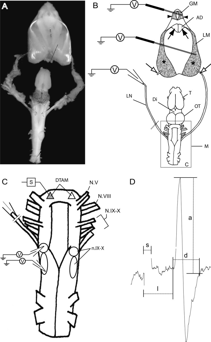
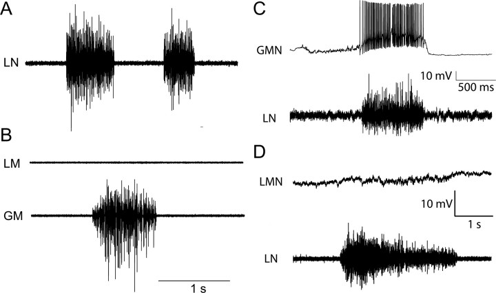
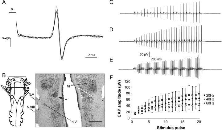
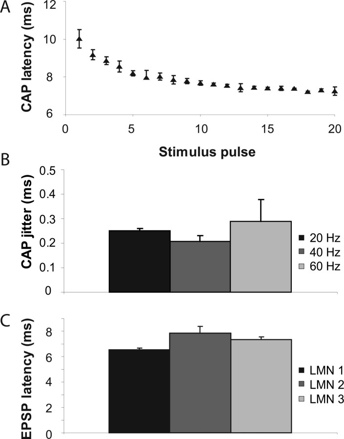
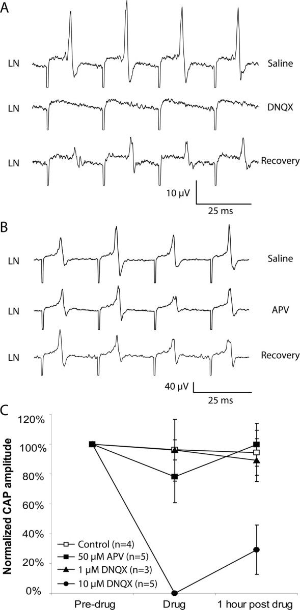
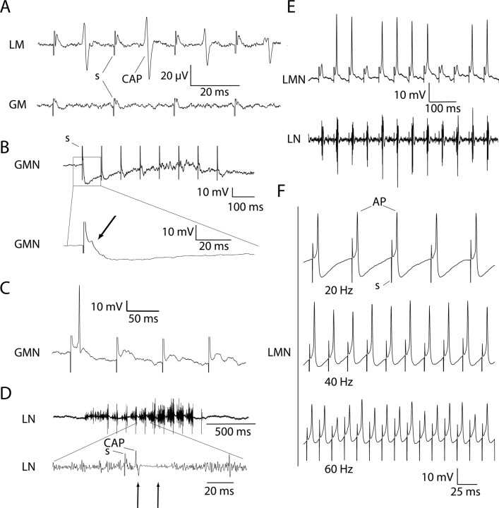
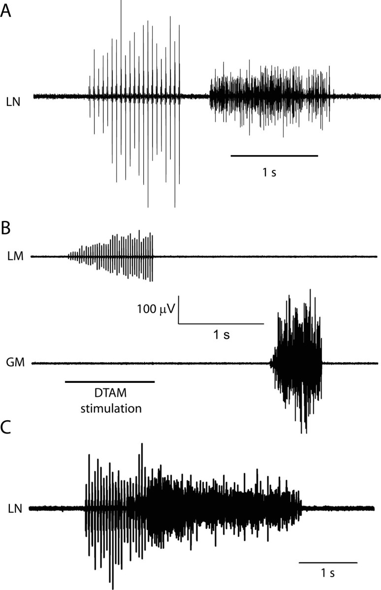
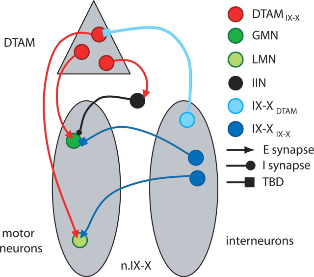
The aquatic frog Xenopus laevis uses a complex vocal repertoire during mating and male-male interactions. Calls are produced without breathing, allowing the frogs to vocalize for long periods underwater. The Xenopus vocal organ, the larynx, is innervated by neurons in cranial motor nucleus (n.) IX-X, which contains both vocal (laryngeal) and respiratory (glottal) motor neurons. The primary descending input to n.IX-X comes from the pretrigeminal nucleus of the dorsal tegmental area of the medulla (DTAM), located in the rostral hindbrain. We wanted to characterize premotor inputs to respiratory and vocal motor neurons and to determine what mechanisms might be involved in regulating two temporally distinct rhythmic behaviors: breathing and calling. Using isolated brain and larynx preparations, we recorded extracellular activity from the laryngeal nerve and muscles and intracellular activity in laryngeal and glottal motor neurons. Spontaneous nerve activities mimicking respiratory and vocal patterns were observed. DTAM projection neurons (DTAM(IX-X) neurons) provide direct input to glottal and laryngeal motor neurons. Electrical stimulation produced short-latency coordinated activity in the laryngeal nerve. DTAM(IX-X) neurons provide excitatory monosynaptic inputs to laryngeal motor neurons and mixed excitatory and inhibitory inputs to glottal motor neurons. DTAM stimulation also produced a delayed burst of glottal motor neuron activity. Together, our data suggest that neurons in DTAM produce vocal motor output by directly activating laryngeal motor neurons and that DTAM may coordinate vocal and respiratory motor activity.


Figures

Figure 1.
Figure 1.
Experimental design: isolated vocal apparatus and physiological recordings. A, Photograph of the isolated “intact” vocal preparation: the brain and larynx. The larynx is pinned anterior to the brain (anterior is up). B, Schematic of the vocal apparatus and extracellular electrode configuration. Laryngeal dilator muscles (*) contract to separate arytenoid cartilage discs (filled arrows). The glottis, anterior to the discs, closed in the relaxed state, is opened by contraction of the glottal muscles on either side (arrowheads). The laryngeal nerve exits the brain via N.IX–X and inserts into the dorsolateral posterior extent of the laryngeal dilators (open arrows). A suction electrode records activity from the cut end of the (left) laryngeal nerve, or the activity of the glottal and laryngeal dilator muscles are recorded directly (right muscles in diagram) when the laryngeal nerve is left intact. C, Detailed diagram of the medulla showing stimulation and intracellular recording sites. Activity in one laryngeal nerve is recorded (left) while the ipsilateral DTAM is stimulated (shaded). Intracellular recordings in n.IX–X were performed ipsilateral to DTAM stimulation sites. D, Example of a typical CAP produced by stimulation. Time between onset of the stimulus artifact (s) and the final upward inflection point before exceeding threshold is measured for CAP latencies (l). CAP duration (d) is measured between onset and the point at which the trace first returns to baseline. Amplitude (a) is measured as maximum deflection from baseline. AD, Arytenoid cartilage disc; Di, diencephalon; GM, glottal muscle; LM, laryngeal dilator muscle; LN, laryngeal nerve; M, medulla; OT, optic tectum; N.V, trigeminal nerve; N.VIII, auditory nerve; N.IX–X, glossolaryngeal–vagal nerve; T, telencephalon. Brain and larynx diagrams adapted from Zornik and Kelley (2007).
Figure 2.
Figure 2.
Spontaneous activity in the isolated brain. A, Recordings from the LN show spontaneous high-frequency, unpatterned bursts. B, In the intact vocal preparation, spontaneous burst activity can be recorded from the glottal (GM), but not laryngeal (LM), muscles. C, Glottal motor neurons (GMN, top trace) rapidly spike during spontaneous glottal bursts on the nerve (bottom trace). D, LMNs (top trace) do not spike during glottal bursts (bottom trace).
Figure 3.
Figure 3.
A, Doublet CAPs (resembling the Xenopus amplectant call pattern) are produced spontaneously by the isolated brain. B, Histogram showing the distribution of amplectant call click intervals (n = 6 animals) and spontaneous CAP intervals (n = 6 preparations) (data are only presented for the first interval; see Results).
Figure 4.
Figure 4.
DTAM stimulation: nerve recordings. A, Repeated DTAM stimulation produces coordinated, stereotyped CAPs (5 overlaid traces are shown). B, Diagram of the medulla; shaded triangle outlines DTAM stimulation site. Box indicates area shown in adjacent photomicrograph. Nissl-stained horizontal section shows lesion (arrow) centered in the left DTAM. DTAM (outlined on the right side) is medial and caudal to nucleus isthmi (NI) and anteromedial to the motor nucleus of the trigeminal (n.V). C, DTAM stimulation at 20 Hz produces a potentiating series of CAPs. D, E, The 40 Hz (D) and 60 Hz (E) stimulation also elicits amplitude modulated CAPs. F, Means of first 20 CAPs during DTAM stimulus trains (quantification of 3 representative preparations). The 40 Hz trains elicit the greatest increase in CAP size; stimuli at 60 Hz elicit the least amount of amplitude modulation. Traces represent an average of five stimulus sweeps.
Figure 5.
Figure 5.
DTAM stimulation: CAP and EPSP latency and jitter. A, Average CAP onset latencies during DTAM stimulation (20 Hz) in one representative brain; latencies decrease as stimulus trains progress. B, CAP jitter values are small and do not vary across DTAM stimulus rates. C, DTAM-induced EPSPs in LMNs have latencies from 6 to 8 ms. Jitter values (SD of the mean; error bars) are small.
Figure 6.
Figure 6.
Premotor stimulation: identification of the presynaptic transmitter. A, Short-latency CAPs produced in response to DTAM stimulation (top trace) are eliminated in the presence of 10 μm bath-applied DNQX (middle trace). Recovery can be achieved after >1 h washout (bottom trace). B, DTAM-induced CAPs (top trace) are not reduced by bath application of 50 μm APV (middle trace). C, Chart summarizing effects of APV and DNQX application to DTAM stimulation-induced CAPs. Values are normalized to pretreatment amplitudes (left column) and expressed as percentage of that value during drug application (middle column) and after washout (right column). All five 10 μm DNQX trials completely eliminated CAP production (black circle, center). LN, Laryngeal nerve.
Figure 7.
Figure 7.
Distinct inputs from DTAM to laryngeal and glottal motor neurons. A, Recordings from laryngeal dilator muscle (LM) and glottal muscle (GM) during DTAM stimulation show EMG potentials recorded from laryngeal dilator muscle but not glottal muscle, suggesting that DTAM-induced CAPs in the nerve represent laryngeal motor neuron activity. B, During stimulation of DTAM, glottal motor neurons (GMNs) are inhibited (top trace) but receive mixed excitatory and inhibitory inputs. The bottom, expanded trace shows an example of an EPSP (arrow) truncated by inhibitory input. C, In some cases, glottal motor neuron EPSPs produced by DTAM stimulation lead to a spike, usually at the beginning of a train. D, Stimulating DTAM during a spontaneous glottal burst results in brief pauses in activity after each stimulus pulse (top trace). Enlarged view of a single DTAM stimulation pulse (bottom trace) shows a resulting CAP and the subsequent pause in glottal burst activity (arrows). E, Intracellular recordings from LMNs (top trace) during DTAM stimulation shows a single EPSP or spike after each pulse, coming shortly before a CAP in the nerve (bottom trace). F, One-to-one action potentials (AP) are induced by 20, 40, and 60 Hz DTAM stimulation.
Figure 8.
Figure 8.
DTAM stimulation induces glottal bursts. A, After stimulation in DTAM, high-frequency unpatterned bursts of activity, recorded from N.IX–X, often follow laryngeal–muscle-associated CAPs. The durations of these unpatterned bursts are similar to spontaneous glottal bursts (Fig. 2A,B). B, EMG recordings from laryngeal (top trace) and glottal (bottom trace) muscles. Top trace shows stimulus-locked CAPs in laryngeal dilator muscle (LM) but not glottal muscle (GM); poststimulation activity is only present in glottal muscle. C, The glottal burst delay after DTAM stimulation requires GABA transmission; bath application of bicuculline (1 μm) eliminates or drastically reduces this delay (n = 5). LN, Laryngeal nerve.
Figure 9.
Figure 9.
Updated model of the Xenopus laevis vocal–respiratory hindbrain circuit. Graphical representations of DTAM (gray triangle, shown unilaterally) and n.IX–X (gray ovals, shown bilaterally). For clarity, only motor neurons are shown in the left n.IX–X, and only interneurons are illustrated in right n.IX–X; in reality, all neuronal pools exist bilaterally. Motor and premotor neurons of the Xenopus vocal–respiratory circuit have been described previously (Zornik and Kelley, 2007). Here, we have shown that DTAMIX–X neurons provide excitatory inputs to both LMNs and glottal motor neurons (GMNs). Our data also indicate the existence of an inhibitory interneuron (IIN; black) that is activated by DTAM; its location (likely in either n.IX–X or DTAM) is as yet uncertain. Another study showed that commissural IX–XIX–X neurons also provide excitatory (APMA receptor mediated) inputs onto LMNs (Zornik, 2006), suggesting that LMNs can be activated by at least two premotor neuron pools. The valence and transmitters of other neurons in the circuit (including IX–XDTAM neurons and IX–XIX–X neurons targeting glottal motor neurons) remain to be determined. E, Excitatory; I, inhibitory; TBD, to be determined.

References

    1. Bass AH, Baker R. Phenotypic specification of hindbrain rhombomeres and the origins of rhythmic circuits in vertebrates. Brain Behav Evol. 1997;50(Suppl 1):3–16. - PMC - PubMed
    1. Brahic CJ, Kelley DB. Vocal circuitry in Xenopus laevis: telencephalon to laryngeal motor neurons. J Comp Neurol. 2003;464:115–130. - PMC - PubMed
    1. Doyle MW, Bailey TW, Jin YH, Andresen MC. Vanilloid receptors presynaptically modulate cranial visceral afferent synaptic transmission in nucleus tractus solitarius. J Neurosci. 2002;22:8222–8229. - PMC - PubMed
    1. Evans BJ, Kelley DB, Tinsley RC, Melnick DJ, Cannatella DC. A mitochondrial DNA phylogeny of African clawed frogs: phylogeography and implications for polyploid evolution. Mol Phylogenet Evol. 2004;33:197–213. - PubMed
    1. Feldmeyer D, Lubke J, Silver RA, Sakmann B. Synaptic connections between layer 4 spiny neurone-layer 2/3 pyramidal cell pairs in juvenile rat barrel cortex: physiology and anatomy of interlaminar signalling within a cortical column. J Physiol (Lond) 2002;538:803–822. - PMC - PubMed

Publication types

MeSH terms

LinkOut - more resources