. 2008 Jan 16;28(3):672-80.
doi: 10.1523/JNEUROSCI.2132-07.2008.

Antiapoptotic and trophic effects of dominant-negative forms of dual leucine zipper kinase in dopamine neurons of the substantia nigra in vivo

Affiliations

Antiapoptotic and trophic effects of dominant-negative forms of dual leucine zipper kinase in dopamine neurons of the substantia nigra in vivo

Xiqun Chen et al. J Neurosci. .

Abstract

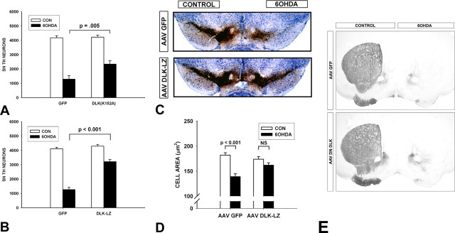
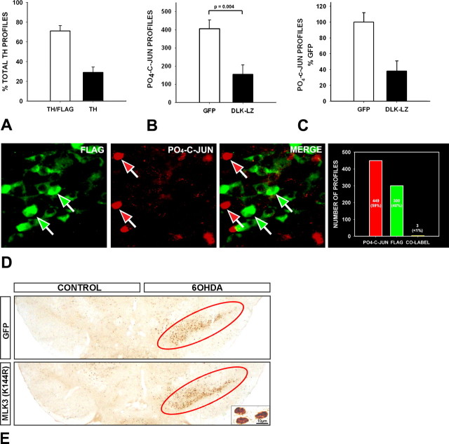
There is extensive evidence that the mitogen-activated protein kinase (MAPK) signaling cascade mediates programmed cell death in neurons. However, current evidence that the mixed linage kinases (MLKs), upstream in this cascade, mediate cell death is based, in the in vivo context, entirely on pharmacological approaches. The compounds used in these studies have neither complete specificity nor selectivity among these kinases. Therefore, to better address the molecular specificity of the MLKs in mediating neuron death, we used dominant-negative constructs delivered by AAV (adenoassociated virus) vector transfer. We assessed effects in a neurotoxin model of parkinsonism, in which neuroprotection by pharmacologic MLK inhibition has been reported. We find that two dominant-negative forms of dual leucine zipper kinase (DLK) inhibit apoptosis and enhance long-term survival of dopamine neurons, but a dominant negative of MLK3 does not. Interestingly, the kinase-dead form of DLK not only blocks apoptosis but also has trophic effects on dopamine neurons. Although the MAPK cascade activates a number of downstream cell death mediators, we find that inhibition of DLK correlates closely with blockade of phosphorylation of c-jun and prevention of cell death. We conclude that DLK acts primarily through c-jun phosphorylation to mediate cell death in this model.


Figures

Figure 1.
Figure 1.
Transduction of DA neurons of the SN with AAV vectors expressing dominant-negative forms of MLKs. The top row of panels illustrates immunoperoxidase labeling (brown) for the FLAG epitope of each transgene in the SN. Positive immunoperoxidase labeling in the SNpc (arrows) is shown for each of the dominant-negative constructs. The following rows of panels demonstrate successful transduction of DA neurons within SNpc for SN-MLK3(K144R), DN-DL (K152A), and DN-DLK-LZ, respectively. The dopaminergic phenotype is demonstrated by immunofluorescent staining for TH (red); transgene expression is demonstrated by staining for the FLAG epitope (green); coexpression is demonstrated by yellow in the merged images in the right-hand panels. Scale bars: 10 μm.
Figure 2.
Figure 2.
Trophic effects of kinase-dead DN-DLK(K152A) on DA neurons of the SN. A, Stereological analysis of the number of TH-positive neurons in the SN at 28 d after AAV injection reveals a 19% increase in comparison with the contralateral noninjected control side (CON) (p < 0.001, ANOVA) (n = 8). No difference in number between the two sides was noted for mice injected with AAV DN-MLK3(K144R) (n = 8) or AAV DN-DLK-LZ (data not shown). B, The mean cross-sectional area of SN DA neurons was increased by 34% in comparison with contralateral controls at 28 d after injection in mice receiving AAV DN-DLK(K152A) (p < 0.001). No significant effect was observed in mice injected with AAV GFP. A total of 50 neurons (25 each in 2 mice) was examined for each condition. C, No effect was observed on the number of NeuN-stained neurons in the SNpc at 28 d after injection of either AAV GFP (n = 6) or AAV DN-DLK(K152A) (n = 5). This result suggests that the increased apparent number of TH-positive neurons in A is attributable to an increased expression of TH, rather than an actual increase in the number of neurons. Error bars indicate SEM. D, A modest (16%) but significant (p < 0.03, paired t test) increase in TH activity (picomoles of 14CO2 formed per minute per milligram of protein) is observed in SN on the side of AAV DN-DLK(K153A) compared with the contralateral control at 28 d after injection (n = 8). A nonsignificant trend (p = 0.3) is observed in the AAV GFP-injected mice (n = 8).
Figure 3.
Figure 3.
Dominant-negative forms of DLK inhibit apoptosis in SN DA neurons after intrastriatal 6OHDA. A, Adult male mice were injected with either AAV GFP or AAV DN-DLK(K152A) and then with 6OHDA 3 weeks later. At 6 d after lesion, animals were killed, and the number of apoptotic profiles in the SNpc was determined. AAV DN-DLK(K152A) reduced the number of apoptotic profiles to 12% of the number observed in AAV GFP controls [p < 0.001, t test; n = 8 GFP controls; n = 7 DN-DLK(K152A)]. B, In a similar experiment, AAV DN-DLK-LZ (n = 7) reduced the number of apoptotic profiles to 18% of that observed in AAV GFP controls (n = 7) (p = 0.02, t test). C, AAV DN-MLK3(K144R) had no effect on the number of apoptotic profiles after 6OHDA. D, To directly compare kinase-dead DN-DLK(K152A) to DN-MLK3(K154R), the viral stocks of AAV DN-DLK(K152A) were diluted to a titer equal to that of AAV DN-MLK3(K144R) (2.4E12 vg/ml). Even at this diluted titer, AAV DN-DLK3(K152A) retained the ability to suppress apoptosis (p = 0.01, t test; n = 11). Error bars indicate SEM.
Figure 4.
Figure 4.
Dominant-negative forms of DLK enhance the long-term survival of SN DA neurons after intrastriatal 6OHDA. A, At 3 weeks after intranigral injection of AAV, mice received intrastriatal 6OHDA. At 28 d after 6OHDA, mice were killed and processed for immunoperoxidase staining of TH within SN neurons and axons. At this postlesion time point, there is no suppression of TH phenotype, and SN DA neuron counts determined by TH staining are approximately equal to those determined by Fluorogold retrograde labeling (Sauer and Oertel, 1994). AAV DN-DLK(K152A) increased the number of surviving DA neurons to 55% of the contralateral control (CON) noninjected side, which represents an 80% increase over the number of surviving neurons in the AAV GFP group [p = 0.005, ANOVA; n = 8 and 7, AAV GFP and AAV DN-DLK(K152A), respectively]. B, In a similar experiment, AAV DLK-LZ increased the number of surviving DA neurons to 75% of the contralateral control, which represents a 153% increase over the number of surviving neurons in the AAV GFP group (p < 0.001, ANOVA; n = 6 and 8 AAV GFP and AAV DN-DLK-LZ, respectively). C, A representative set of sections stained for TH (brown) and Nissl-counterstained after injection of AAVs and unilateral intrastriatal 6OHDA. At 28 d after 6OHDA, a mouse given AAV GFP shows very few surviving DA neurons in the SNpc. A mouse given AAV DN-DLK-LZ shows substantial preservation of DA neurons. D, AAV DN-DLZ-LZ preserves DA neuron size after 6OHDA. In the AAV GFP control condition, there is a 24% loss of cross-sectional area (p < 0.001, ANOVA; n = 100 neurons), whereas in the AAV DN-DLK-LZ condition there is only a 7% trend (p = 0.4, NS). Error bars indicate SEM. E, Neither AAV DN-DLK(K152A) nor AAV DN-DLK-LZ protected dopaminergic axons. Representative TH-immunostained striatal sections demonstrate that, for both GFP (top) and AAV DLK-LZ (bottom), there is near-complete loss of striatal TH staining after intrastriatal 6OHDA. Similar results were observed with AAV DN-DLK(K152A) (data not shown).
Figure 5.
Figure 5.
Transduction with AAV DN-DLK-LZ blocks phosphorylation of c-jun. A, To determine the transduction efficiency of AAV DN-DLK-LZ, n = 4 mice were injected into the SN and killed 4 weeks later for double-labeling of TH and FLAG. In sections from each animal, n = 25 TH-positive profiles (n = 100 total) were assessed for FLAG staining; 71.1 ± 6% of TH profiles were FLAG-positive. B, C, The efficacy of AAV DN-DLK-LZ to suppress c-jun phosphorylation was assessed at the population level, by counting the number of PO4-c-jun-positive immunostained profiles at 6 d after 6OHDA, in mice which received either AAV GFP (n = 7) or AAV DN-DLK-LZ (n = 10). Absolute counts are shown in B; counts as a percentage of the mean among the AAV GFP mice is shown in C. AAV DN-DLK-LZ reduced the number of PO4-c-jun profiles to 38 ± 13% of the number observed in AAV GFP mice. Error bars indicate SEM. D, The relationship between transduction with AAV DN-DLK-LZ and phosphorylation of c-jun after 6OHDA was examined at the cellular level by double-labeling for FLAG and PO4-c-jun at 6 d after 6OHDA. Of 752 profiles examined in sections from three mice, 449 (59.7%) stained for PO4-c-jun alone, 300 (39.8%) stained for FLAG alone, and only 3 profiles (0.4%) stained for both. Thus, phosphorylation of c-jun in the presence of AAV DN-DLK-LZ was exceedingly rare. A representative field demonstrates the discordance between FLAG and PO4-c-jun staining at the cellular level. E, Transduction of SNpc neurons with AAV DN-MLK3(K144R) does not block phosphorylation of c-jun after 6OHDA. The abundance of PO4-c-jun-positive nuclear profiles is about the same after AAV DN-MLK3(K144R) as it is after AAV GFP, as shown in a representative low-power micrograph in E and confirmed by quantitative analysis (data not shown). Representative phospho-c-jun nuclear staining is shown at high magnification in the inset in E. Positive phospho-c-jun staining in the GFP and MLK3(K144R) conditions, on the side of the 6OHDA injection, is shown in the low-power micrographs encircled in red.

References

    1. Bradford MM. A rapid and sensitive method for the quantitation of microgram quantities of protein utilizing the principle of protein-dye binding. Anal Biochem. 1976;72:248–254. - PubMed
    1. Burke RE. Inhibition of MAPK and stimulation of Akt kinase signaling pathways: two approaches with therapeutic potential in the treatment of neurodegenerative disease. Pharmacol Ther. 2007;114:261–277. - PMC - PubMed
    1. Coleman M. Axon degeneration mechanisms: commonality amid diversity. Nat Rev Neurosci. 2005;6:889–898. - PubMed
    1. Crocker SJ, Lamba WR, Smith PD, Callaghan SM, Slack RS, Anisman H, Park DS. c-Jun mediates axotomy-induced dopamine neuron death in vivo. Proc Natl Acad Sci USA. 2001;98:13385–13390. - PMC - PubMed
    1. Donovan N, Becker EB, Konishi Y, Bonni A. JNK phosphorylation and activation of BAD couples the stress-activated signaling pathway to the cell death machinery. J Biol Chem. 2002;277:40944–40949. - PubMed

Publication types

MeSH terms

LinkOut - more resources