. 2008 Jan 16;28(3):696-710.
doi: 10.1523/JNEUROSCI.4931-07.2008.

Intrinsic mechanisms for adaptive gain rescaling in barrel cortex

Affiliations

Intrinsic mechanisms for adaptive gain rescaling in barrel cortex

Marta Díaz-Quesada et al. J Neurosci. .

Abstract

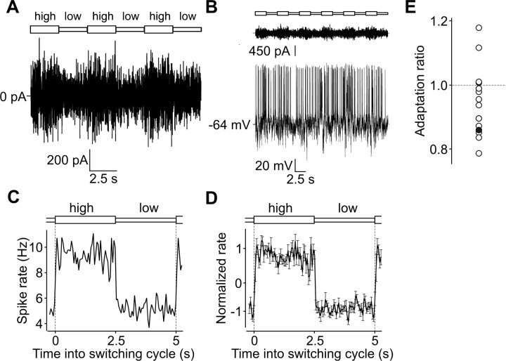
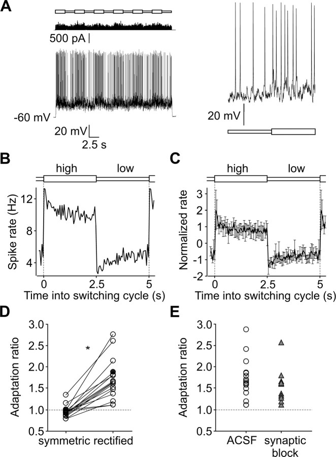
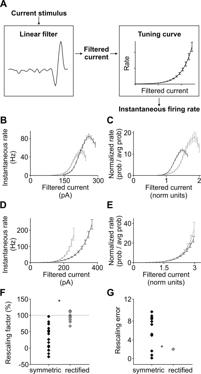
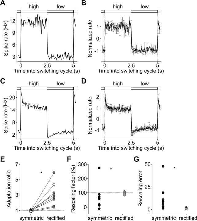
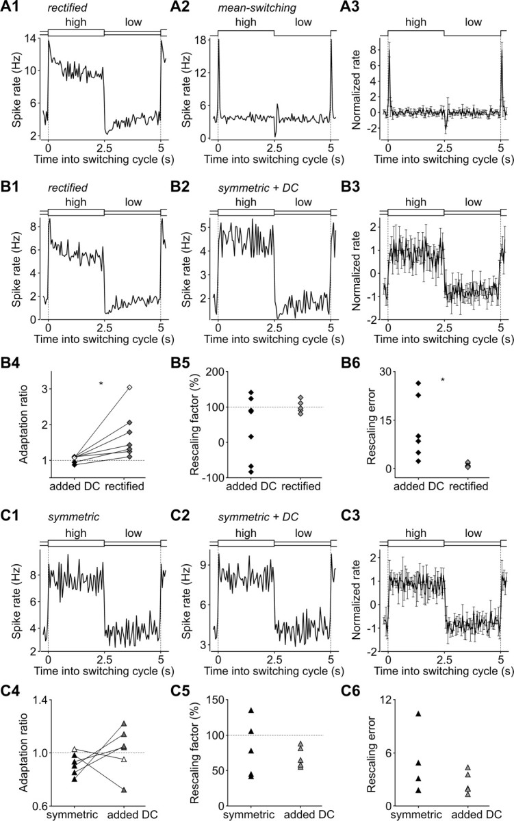
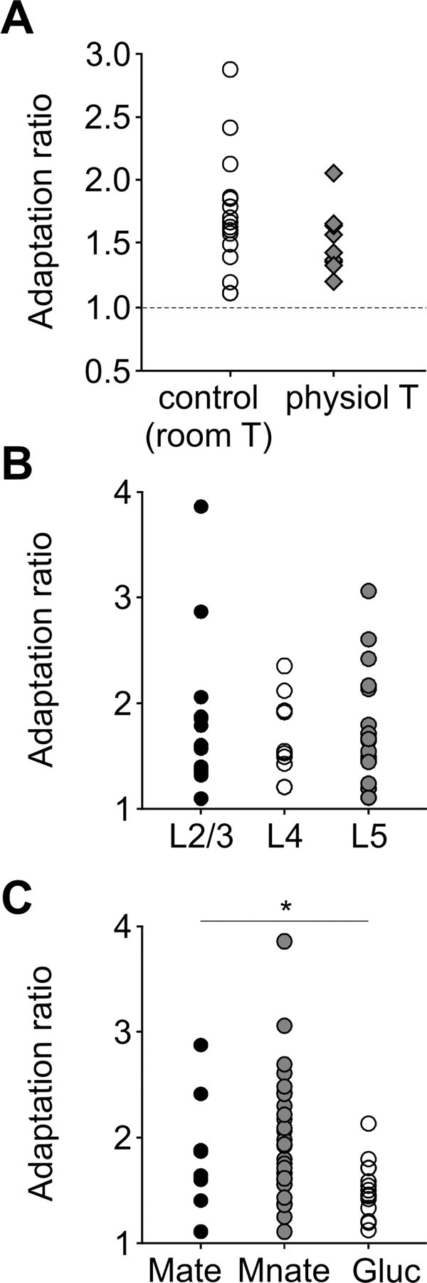
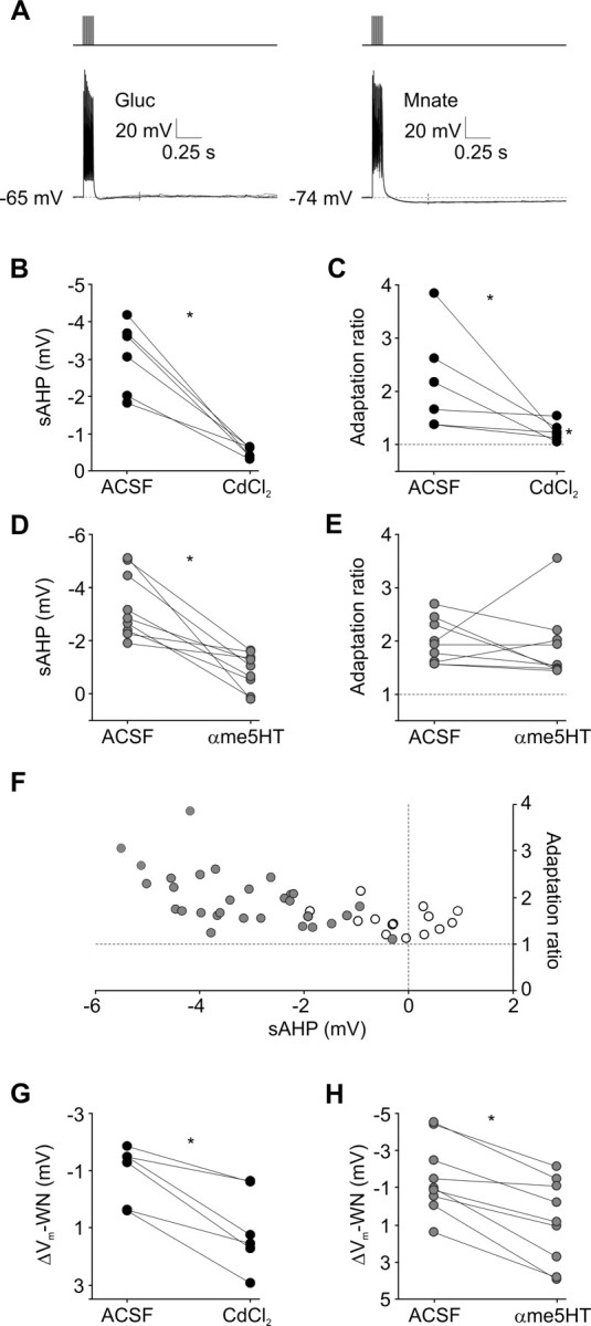
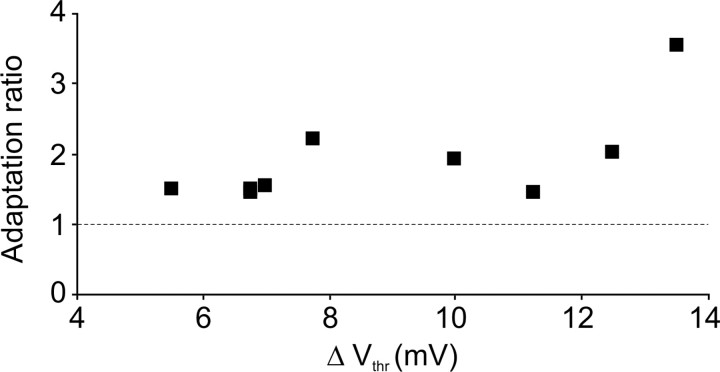
Barrel cortex neuronal responses adapt to changes in the statistics of complex whisker stimuli. This form of adaptation involves an adjustment in the input-output tuning functions of the neurons, such that their gain rescales depending on the range of the current stimulus distribution. Similar phenomena have been observed in other sensory systems, suggesting that adaptive adjustment of responses to ongoing stimulus statistics is an important principle of sensory function. In other systems, adaptation and gain rescaling can depend on intrinsic properties; however, in barrel cortex, whether intrinsic mechanisms can contribute to adaptation to stimulus statistics is unknown. To examine this, we performed whole-cell patch-clamp recordings of pyramidal cells in acute slices while injecting stochastic current stimuli. We induced changes in statistical context by switching across stimulus distributions. The firing rates of neurons adapted in response to changes in stimulus statistics. Adaptation depended on the form of the changes in stimulus distribution: in vivo-like adaptation occurred only for rectified stimuli that maintained neurons in a persistent state of net depolarization. Under these conditions, neurons rescaled the gain of their input-output functions according to the scale of the stimulus distribution, as observed in vivo. This stimulus-specific adaptation was caused by intrinsic properties and correlated strongly with the amplitude of calcium-dependent slow afterhyperpolarizations. Our results suggest that widely expressed intrinsic mechanisms participate in barrel cortex adaptation but that their recruitment is highly stimulus specific.


Figures

Figure 1.
Figure 1.
Absence of adaptation to stimulus statistics under symmetric current stimuli in barrel cortex. A, Stimulus segment showing three 5 s cycles of high/low variance switching. High- and low-variance epochs are represented in the top diagram. Stimulus was distributed as a Gaussian and was symmetric about its 0 pA mean. B, High- and low-variance epochs (diagram as for A; top trace), stimulus (current; middle trace), and response (membrane potential; bottom trace) for a typical neuron. Traces depict a complete trial. Numerical value at left indicates resting membrane potential. The neuron fired strongly during high-variance epochs and more weakly during low-variance epochs. C, Firing rate plot for same neuron as B, showing an absence of rate adaptation. The plot was constructed by binning spike times within each cycle (using 50 ms windows) and averaging responses over the number of cycles in the experiment. High- and low-variance epochs are represented in the top diagram; the transition from low to high is shown at both edges to emphasize the cyclic character of the plot. D, Population pooled firing rate plot, showing normalized firing rate averaged over neurons (n = 16). Error bars represent 1 SD. Adaptation was absent across the population. E, Adaptation ratio plot (see Materials and Methods). Dark symbol depicts neuron in B and C. Adaptation ratios >1 signified adaptation; in this population, ratios were somewhat, but not significantly, <1.
Figure 2.
Figure 2.
Adaptation to stimulus statistics under rectified stimuli. A, Left, High- and low-variance epochs (top), stimulus (current; middle), and response (membrane potential; bottom) for rectified stimuli, for a different neuron to that in Figure 1B. Right, Magnified membrane potential response, −80 to +80 ms relative to a low- to high-variance transition. Bottom diagram indicates low- and high-variance periods. Length of bars, 80 ms. Note tendency toward higher firing threshold during high-variance portion of trace. B, Firing rate plot for same neuron as A, constructed as Figure 1C, showing clear adaptation to rectified stimuli. C, Population rate plot (n = 16) showing significant adaptation to rectified stimuli. Error bars represent 1 SD. D, Adaptation ratio plot comparing ratio values across symmetric and rectified conditions. Lines connect symmetric and rectified data points for each neuron (n = 16). Asterisk denotes significant difference; also, ratios for rectified stimuli were significantly >1. Dark symbol depicts neuron in A and B. E, Adaptation was unchanged in the presence of synaptic blockers. Symbols and colors arbitrarily chosen to aid discrimination of data points.
Figure 3.
Figure 3.
Full gain rescaling occurred only for rectified stimuli. A, Schematic of LN framework for characterizing neuronal stimulus–response relationships. Examples of linear filter and tuning curve correspond to the same neuron as the panels below, tested with rectified stimuli. B, Absolute input–output functions, or tuning curves, for neuron in Figure 2A tested with symmetric stimuli. Curves were computed separately for high-variance epoch (dark line) and low-variance epoch (light line). Output is the predicted instantaneous firing probability in units of rate; input is the current stimulus projected onto the most significant neuronal filter from spike-triggered analysis (see Materials and Methods). Error bars represent SD from 30 repetitions of the estimation procedure. Note slight rescaling along x-axis: over values up to ∼200 pA, more current was necessary to reach a given firing rate in the high-variance epoch. The curve folds over for higher values. C, Normalized input–output functions for same recording as in B. Output is normalized to the average rate, thus representing how the input modulates the spiking probability of the neuron relative to its average; input is normalized by the SD of the current distribution. Rescaling is clearly not complete. D, Absolute input–output functions for same neuron as in B and C tested with rectified stimuli. The width of the curves depended more strongly on the width of the distribution; in the high-variance epoch, significantly more current was required to achieve a given rate. E, Normalized input–output functions, computed as in C, show complete rescaling. F, Rescaling factors across the same neuronal population as in Figure 2, tested with symmetric and rectified stimuli. For each neuron, the high-variance fitted tuning curve was multiplicatively rescaled along the x-axis until it best resembled the low-variance curve; plot depicts the resulting best rescaling factor, in units such that 100% (dashed line) corresponds to full rescaling and 0% to no rescaling. Factors under rectified stimulation, but not symmetric stimulation, clustered closely around 100%.Here and in G, asterisk denotes significant difference across distributions. G, Error values for rescaling across the population. Error values were computed from the difference between tuning curves after applying the optimal rescaling factors shown in F. Perfect rescaling would give a negligible error. Rectified stimulation, but not symmetric stimulation, gave well constrained errors. Not shown (out of scale) are four outlying values (>30) corresponding to symmetric stimuli.
Figure 4.
Figure 4.
Preservation of adaptation and gain rescaling under rectified stimuli for a different ratio of SDs. A, Firing rate plot for a neuron, constructed as Figure 1, showing absence of adaptation to symmetric stimuli with a low- to high-SD ratio equal to 0.5. B, Population rate plot for symmetric stimuli (n = 8) showing absence of adaptation. Here and in D, error bars represent 1 SD. C, Rate plot for same neuron as A, showing pronounced adaptation to rectified stimuli with a low- to high-SD ratio equal to 0.5. D, Population rate plot for rectified stimuli (n = 8) showing significant adaptation. Also shown (gray line) is the average population rate for neurons tested with a ratio of SDs equal to 0.7. The gray and black lines overlap with each other, demonstrating the similarity of the behavior for different SD ratio parameters. E, Adaptation ratio plot comparing ratio values across symmetric and rectified conditions. Lines connect symmetric and rectified data points for each neuron (n = 8). Asterisk denotes significant difference; also, ratios for rectified stimuli were significantly >1. White symbol depicts neuron in A and C. F, Rescaling factors across population, tested with symmetric and rectified stimuli. Asterisk denotes significant difference across distributions. Rescaling factors computed as in Figure 3F. G, Error values for rescaling across population. Rectified stimulation, but not symmetric stimulation, gave well constrained errors. Errors computed as in Figure 3G.
Figure 5.
Figure 5.
Stimulus dependence of adaptation. A, Comparison between responses to rectified stimuli and mean-switching stimuli. A1, A2, Rate plots for responses to rectified stimuli (A1) and mean-switching stimuli (A2) for one neuron. Plots constructed as in Figure 1C. Note the striking difference in rate adaptation characteristics, despite the similar firing rate reached at steady state during the “low” epoch. A3, Population rate plot for mean-switching data (n = 8). Error bars depict 1 SD. B, Comparison between responses to rectified stimuli and symmetric stimuli with added DC (for explanation, see Results). B1, B2, Rate plots for responses to rectified stimuli (B1) and added DC stimuli (B2) for a neuron different to the one in A1 and A2. Lack of adaptation under added DC stimuli is similar to behavior under symmetric stimuli in Figure 1, despite the increased excitation provided by the DC. B3, Population rate plot for added DC data (n = 7). Error bars as in A3. B4, Adaptation ratios across population tested with rectified and added DC stimuli. Lines connect data points for each neuron (n = 7). Asterisk denotes significant difference. White symbol depicts neuron in B1 and B2. B5, Rescaling factors across population tested with rectified and added DC stimuli. Factors computed as in Figure 3F. B6, Error values for rescaling across population tested with rectified and added DC stimuli. Asterisk denotes significant difference across distributions. Errors computed as in Figure 3G. C, Comparison between responses to symmetric stimuli without and with added DC (for explanation, see Results). C1, C2, Rate plots for responses to symmetric stimuli (C1) and added DC stimuli (C2) for a neuron different from those in A1, A2 and B1, B2. Lack of adaptation is similar with or without added DC. C3, Population rate plot for added DC data (n = 6). Error bars as in A3. C4, Adaptation ratios across population tested with symmetric and added DC stimuli. Lines connect data points for each neuron (n = 6). There was no significant difference in adaptation. White symbol depicts neuron in C1 and C2. C5, Rescaling factors across population tested with symmetric and added DC stimuli. Factors computed as in Figure 3F. Adding DC did not significantly change the distribution of rescaling factors. C6, Error values for rescaling across population tested with symmetric and added DC stimuli. Errors computed as in Figure 3G. Adding DC made no difference to the distribution of rescaling errors.
Figure 6.
Figure 6.
Experimental analysis of intrinsic adaptation. For all panels, symbols and colors were arbitrarily chosen to aid discrimination of data points. A, Adaptation was present at physiological temperatures (34 ± 1°C), albeit slightly smaller in magnitude. B, Adaptation was not significantly different across neurons from different cortical layers. C, Adaptation was different across neurons recorded with different internal anions. Mate, Potassium methylsulfate recordings; Mnate, potassium methylsulfonate recordings; Gluc, potassium gluconate recordings. Asterisk denotes significant difference.
Figure 7.
Figure 7.
Strong, but not complete, correlation between adaptation and slow AHP current. For all panels, asterisks denote significant differences. A, Standardized protocol for sAHP measurement: 10 current pulses delivered at 100 Hz (top traces). Voltage traces (bottom) are from a gluconate recording (Gluc; left) and a methylsulfonate recording (Mnate; right). Numerical values indicate resting membrane potential. Methylsulfonate recordings had larger sAHPs, which were measured 400 ms after the stimulus train (thin vertical lines show time point). B, CdCl2 significantly decreased pulse train-evoked sAHPs. Lines connect the data points of each neuron (n = 6). C, CdCl2 significantly decreased adaptation under rectified stimulation, although adaptation ratios were still larger than 1 (right asterisk). D, αme5HT also significantly decreased pulse train-evoked sAHPs (n = 9). E, However, αme5HT did not significantly reduce adaptation. F, Adaptation ratios versus pulse train-evoked sAHPs. Gray symbols, Methylsulfonate recordings; white symbols, gluconate recordings. There was a significant correlation between adaptation ratio and sAHP for methylsulfonate, but not gluconate, recordings. Variations in the sAHP explained 32% of the variance in adaptation. G, CdCl2 significantly decreased the average voltage deflection from baseline that remained 400 ms after the end of white noise stimuli, ΔVm-WN (n = 6). H, αme5HT also significantly decreased ΔVm-WN (n = 9).
Figure 8.
Figure 8.
Weak correlation between adaptation and sodium current inactivation. Plot shows data from responses to rectified stimuli in the presence of αme5HT (n = 9). The adaptation ratio value of each neuron is plotted against the corresponding difference in spike threshold between high- and low-variance epochs (ΔVthr). Although these data showed a correlation between adaptation ratio and ΔVthr, overall data using standard ACSF did not.

Similar articles

Cited by

References

    1. Aguera y Arcas B, Fairhall AL. What causes a neuron to spike? Neural Comput. 2003;15:1789–1807. - PubMed
    1. Aguera y Arcas B, Fairhall AL, Bialek W. Computation in a single neuron: Hodgkin and Huxley revisited. Neural Comput. 2003;15:1715–1749. - PubMed
    1. Ahissar E, Sosnik R, Haidarliu S. Transformation from temporal to rate coding in a somatosensory thalamocortical pathway. Nature. 2000;406:302–306. - PubMed
    1. Ahissar E, Sosnik R, Bagdasarian K, Haidarliu S. Temporal frequency of whisker movement. II. Laminar organization of cortical representations. J Neurophysiol. 2001;86:354–367. - PubMed
    1. Arabzadeh E, Zorzin E, Diamond ME. Neuronal encoding of texture in the whisker sensory pathway. PLoS Biol. 2005;3:e17. - PMC - PubMed

Publication types