. 2008 Jan 16;28(3):711-24.
doi: 10.1523/JNEUROSCI.5283-07.2008.

Kalirin-7 is an essential component of both shaft and spine excitatory synapses in hippocampal interneurons

Affiliations

Kalirin-7 is an essential component of both shaft and spine excitatory synapses in hippocampal interneurons

Xin-Ming Ma et al. J Neurosci. .

Abstract

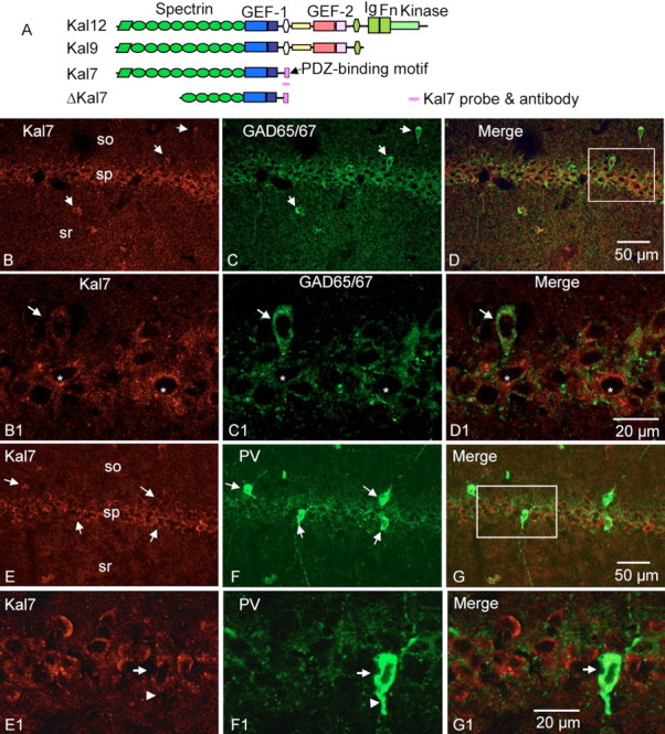
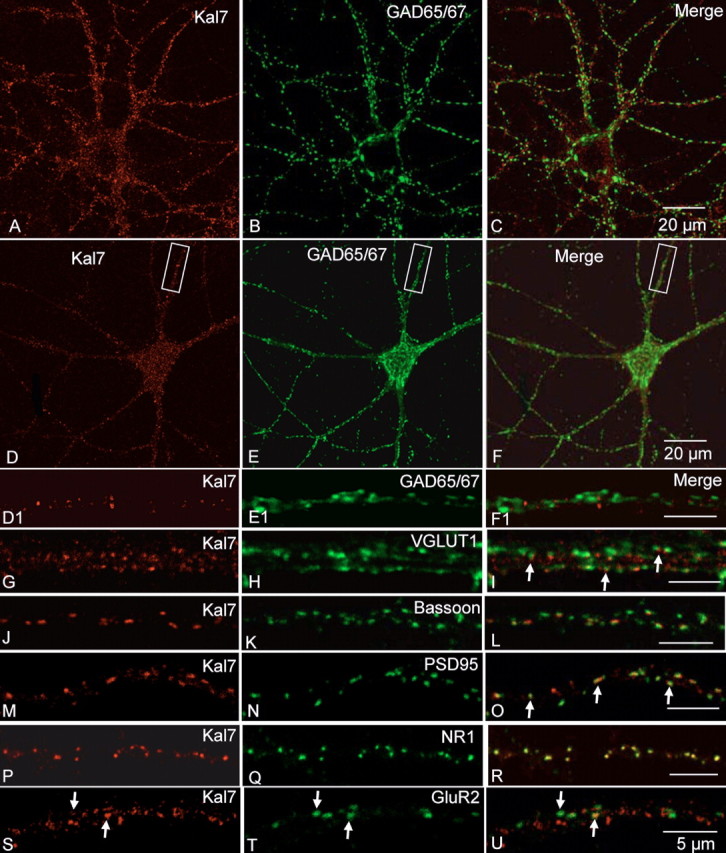
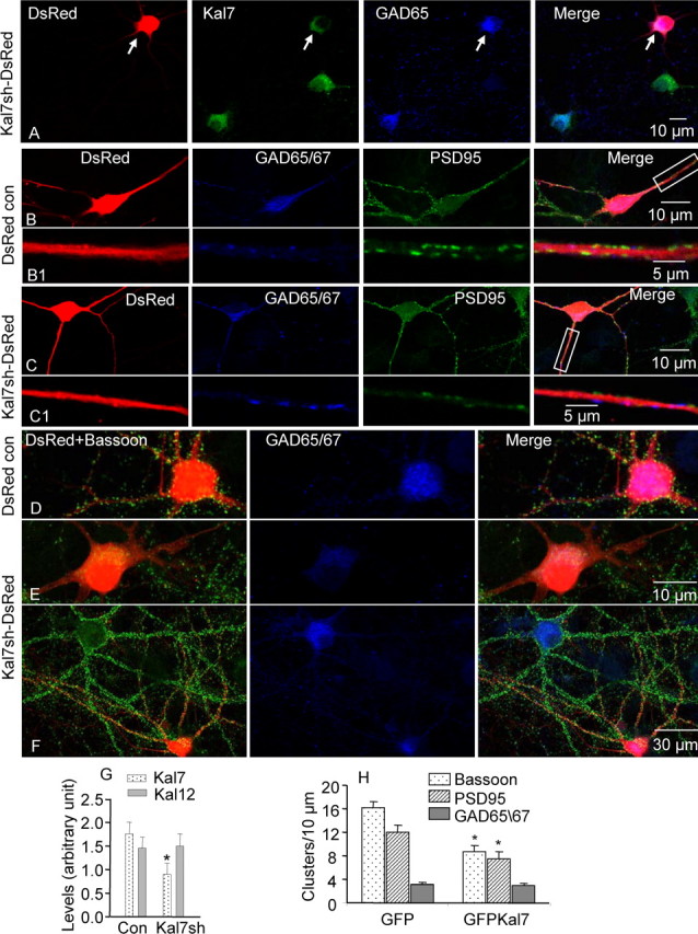
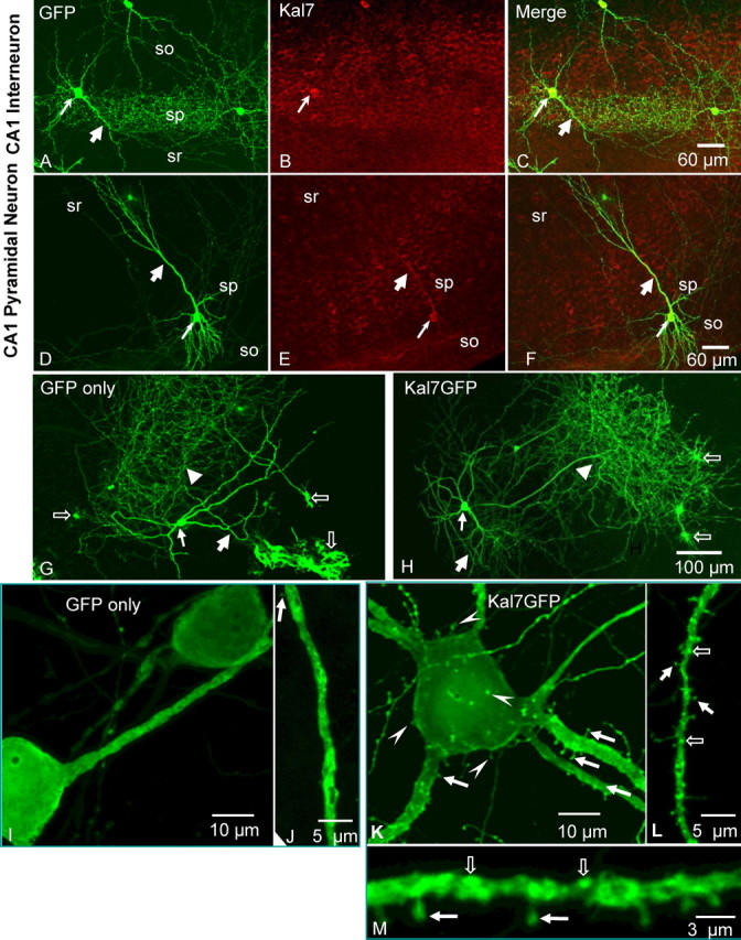
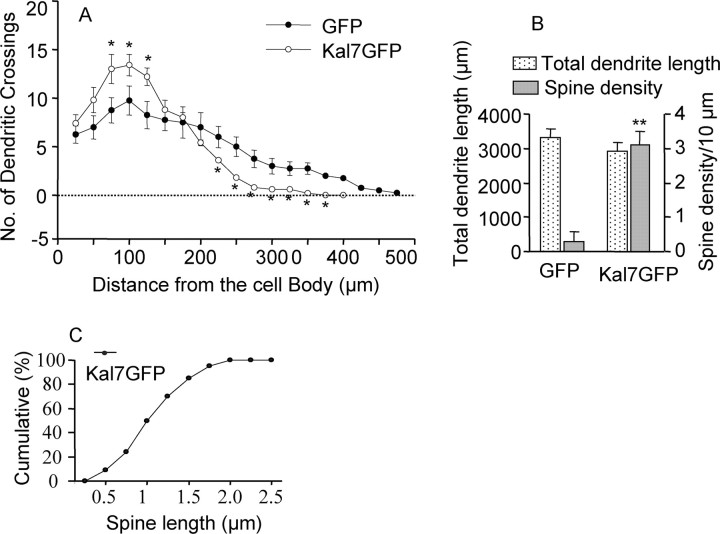
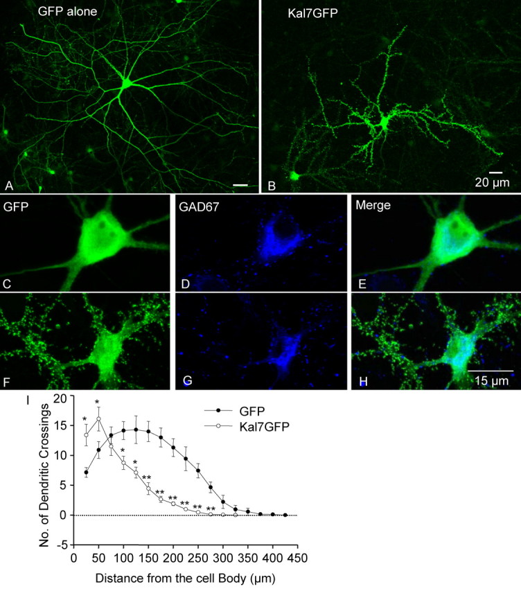
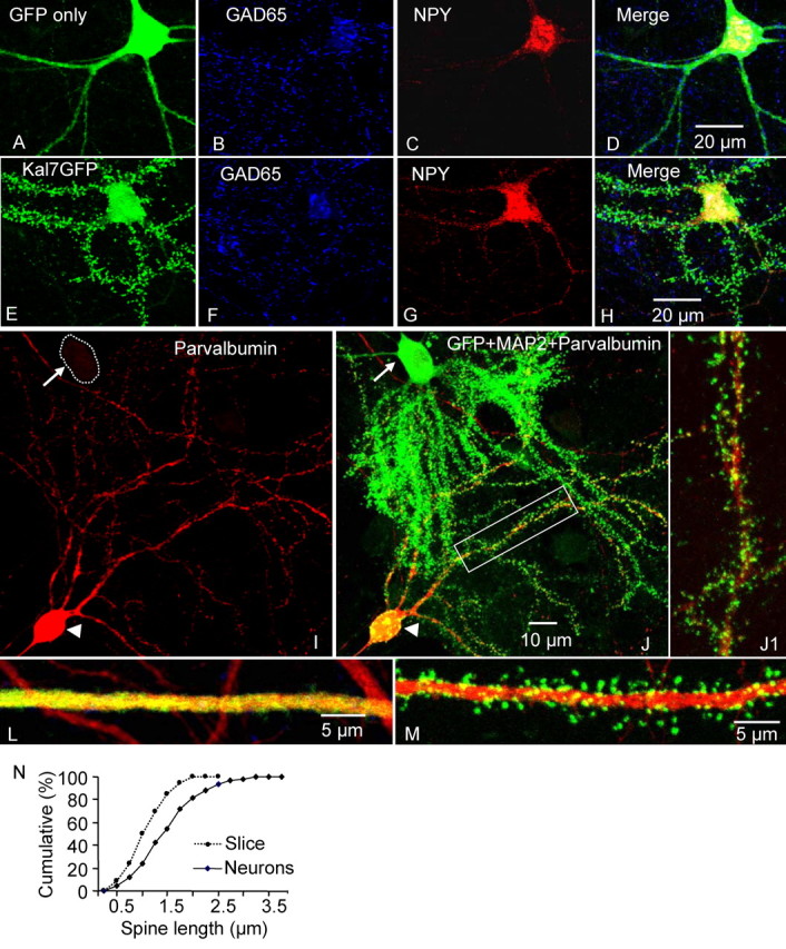
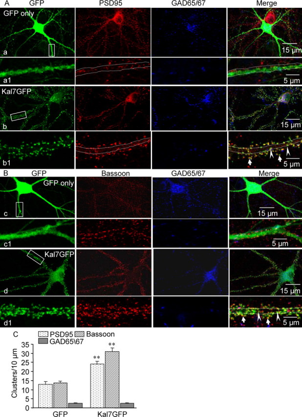
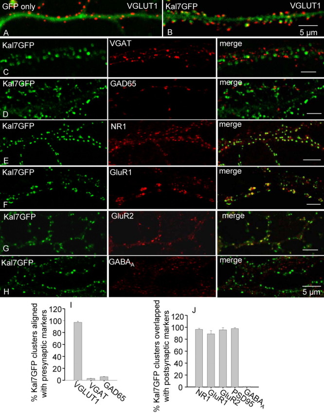
Kalirin, a multifunctional Rho GDP/GTP exchange factor, plays a vital role in cytoskeletal organization, affecting process initiation and outgrowth in neurons. Through alternative splicing, the Kalirin gene generates multiple functionally distinct proteins. Kalirin-7 (Kal7) is the most prevalent isoform in the adult rat hippocampus; it terminates with a postsynaptic density-95 (PSD-95)/Discs large/zona occludens-1 (PDZ) binding motif, is localized to the postsynaptic density, interacts with PSD-95, and causes the formation of dendritic spines when overexpressed in pyramidal neurons. Levels of Kal7 are low in the dendrites of hippocampal aspiny interneurons. In these interneurons, Kal7 is localized to the postsynaptic side of excitatory synapses onto dendritic shafts, overlapping clusters of PSD-95 and NMDA receptor subunit NR1. Selectively decreasing levels of Kal7 decreases the density of PSD-95-positive, bassoon-positive clusters along the dendritic shaft of hippocampal interneurons. Overexpression of Kal7 increases dendritic branching, inducing formation of spine-like structures along the dendrites and on the soma of normally aspiny hippocampal interneurons. Essentially all of the spine-like structures formed in response to Kal7 are apposed to vesicular glutamate transporter 1-positive, bassoon-positive presynaptic endings; GAD-positive, vesicular GABA transporter-positive inhibitory endings are unaffected. Almost every Kal7-positive dendritic cluster contains PSD-95 along with NMDA (NR1) and AMPA (GluR1 and GluR2) receptor subunits. Kal7-induced formation of spine-like structures requires its PDZ binding motif, and interruption of interactions between the PDZ binding motif and its interactors decreases Kal7-induced formation of spine-like structures. Kal7 thus joins Shank3 and GluR2 as molecules with a level of expression at excitatory synapses that titrates the number of dendritic spines.


Figures

Figure 1.
Figure 1.
Kal7 is expressed in hippocampal interneurons. A, The Kalirin gene generates at least 10 transcripts encoding functionally distinct proteins. The largest isoform, Kal12, has multiple spectrin-like repeats, two Rho GEF domains, and a kinase domain. Kal7 has a single GEF domain followed by a PDZ binding motif. The Δ isoforms of Kalirin are generated from a different promoter, and antibodies and probes to the unique region of Kal7 also recognize ΔKal7. B–D, Double immunostaining for Kal7 (B) and GAD65/67 (C) identified Kal7 in interneurons (defined as GAD-positive) in the CA1 region of the adult rat hippocampus. Arrows indicate interneurons in the strata oriens (so) and radiatum (sr) and stratum pyramidal (sp). B1–D1 are high-power images from boxed areas of B–D, respectively. Stars represent pyramidal neurons. E–G, Simultaneous immunostaining for Kal7 (E) and PV (F) identified Kal7 in PV-positive interneurons (arrows) in the CA1 region of the adult rat hippocampus. E1–G1 are high-power images from boxed areas of images E–G, respectively. The dendrite (arrowhead) extending from the soma of the PV-positive neuron does not contain a high level of Kal7.
Figure 2.
Figure 2.
Kal7 is localized at excitatory synapses of hippocampal interneurons. A–F, Double immunostaining of dissociated hippocampal neurons (21 DIV) reveals Kal7 (A, D) in the soma and dendrites of GAD65/67-negative (B) and GAD65/67-positive (E) cells. GAD65/67-positive terminals surround the GAD65/67-negative soma (B). D1–F1, Enlargement of dendrite from the boxed area of D–F shows that Kal7 (D1) is not colocalized with GAD-positive (E1) inhibitory presynaptic terminals (F1, merge). G–U, Dendrites of hippocampal interneurons were stained simultaneously for Kal7 (G, J, M, P, S) and VGLUT1 (H, marker for excitatory presynaptic terminals), Bassoon (K, presynaptic marker), PSD-95 (N, postsynaptic density protein), NMDA receptor (Q, NR1), or AMPA receptor subunit GluR2 (T). Merged images indicate that Kal7 is localized on the postsynaptic side of excitatory synapses. Interneurons were identified by GAD65/67 staining (data not shown). Arrows show the relationship of two synaptic proteins. I, Kal7 clusters were always aligned with VGLUT1 clusters. O, Kal7 clusters were always positive for PSD-95. U, Kal7 clusters were not always positive for CluR2.
Figure 3.
Figure 3.
Expression of Kal7 shRNA causes a reduction in the number of excitatory synapses onto dendritic shafts of interneurons. Cultured hippocampal neurons (14 DIV) were transfected with pSIREN-Kal7 shRNA (A, C, E, F) or pSIREN-DsRed (B, D). A, Cells were stained at 20 DIV with antibodies specific for Kal7 (green) and GAD65 (blue); DsRed marks transfected neurons. Expression of Kal7 shRNA (arrow) caused a decrease in Kal7 staining (A, G; n = 16) and did not alter Kal12 staining (G; n = 14). B, C, Cells were double stained with antibodies specific to GAD65/67 (blue) and PSD-95 (green). B1 and C1 are high-power images of B and C, respectively. Expression of Kal7-shRNA reduced the number of PSD-95-positive clusters in dendrites (C1, H; n = 15). D–F, Cells were double stained with antibodies specific to bassoon (green) and GAD65/67 (blue); DsRed is red. Expression of Kal7-shRNA reduced the number of bassoon-positive presynaptic clusters (H; n = 15). However, the number of GAD65/67-positive clusters along the dendrites was not altered (H; n = 12). Student's t test, *p < 0.05.
Figure 4.
Figure 4.
Expression of Kal7GFP alters the number of dendritic branches and spines in CA1 interneurons. Kal7GFP was expressed in CA1 hippocampal interneurons (A–C) and pyramidal neurons (D–F). Hippocampal slices were biolistically transfected with vector encoding Kal7GFP. Slices fixed 72 h after transfection were immunostained with antibodies to GFP (A, D) and Kal7 (B, E). Interneurons (A–C, thin arrows) were distinguished from CA1 pyramidal neurons (D–F, thin arrows) based on morphology and localization. Thick arrows, Dendrites; so, stratum oriens; sr, stratum radiatum; sp, stratum pyramidal. G, H, Interneurons (thin arrows) and glia (open arrows) from the CA1 area of hippocampal slices were biolistically transfected with vectors encoding GFP (G) or Kal7GFP (H). Thick arrows, Dendrites; arrowheads, axons. I–K, High-power images, showing GFP-positive interneuron is free of spines, and Kal7GFP-positive interneuron contains spines in soma (arrowheads) and dendrite (solid arrows). Some spines are mushroom structures with clear heads (L, M, solid arrows); Kal7GFP-positive clusters are also found within the dendritic shaft (L, M, open arrows).
Figure 5.
Figure 5.
Expression of Kal7GFP alters the number of dendritic branches and spines in CA1 interneurons. A, Sholl analysis (n = 13 for GFP; n = 12 for Kal7GFP). B, Left y-axis, Total dendritic length in CA1 interneurons. B, Right axis, Spine density in CA1 interneurons (GFP, 0.28 ± 0.31/10 μm, n = 13; Kal7GFP, 3.10 ± 0.40/10 μm, n = 12). C, Cumulative frequency plot of spine length in neurons expressing Kal7GFP (800 spines in 12 interneurons). One-way ANOVA followed by Dunnett's test to assess statistical significance between groups (A). Student t test (B), *p < 0.05, **p < 0.01.
Figure 6.
Figure 6.
Expression of Kal7GFP alters dendritic morphology in cultured hippocampal interneurons. Hippocampal neurons in dissociated culture were transfected with vectors encoding GFP (A, C–E) or Kal7GFP (B, F–H) at 1 DIV. Neurons were triple stained at 16 DIV with antibodies to GFP (A, B), MAP2 (data not shown), and GAD65/67 (D, E, G, H). C–E and F–H are high-power images from A and B, respectively. Sholl analysis (I) (n = 17 for GFP; n = 14 for Kal7GFP). One-way ANOVA followed by Dunnett's test to assess statistical significance between groups (I), *p < 0.05, **p < 0.01.
Figure 7.
Figure 7.
Overexpression of Kal7 forces aspiny interneurons to produce spine-like structures. Hippocampal neurons in dissociated culture were transfected with vector encoding GFP alone (A, L) or Kal7GFP (E, J, M) at 1 DIV. A–H, Cultures were triple stained with antibodies to GFP (A, E, green), GAD65 (B, F, red), and NPY (C, G, blue). Merged images are shown in D and H. I, J, Cultures were double stained with antibodies to parvalbumin (I, red) and GFP (green in merged image J). J1 is high-power image from boxed dendrite of J. L, M, Merged images (red and green) from the dendrites of interneurons (see Fig. S4 for lower-power images, available at www.jneurosci.org as supplemental material), which were transfected with GFP alone (L) or Kal7GFP (M), triple stained with antibodies to GFP (green), MAP2 (red), and GAD65/67 (data not shown) (Fig. S4, available at www.jneurosci.org as supplemental material). Spine density in hippocampal interneurons was quantified (0.24 ± 1.1/10 μm for GFP; 15.03 ± 0.70/10 μm for GFPKal7; p < 0.01, Student t test; n = 16). N, Solid line, Cumulative frequency plot of spine length in neurons transfected with Kal7GFP (1000 spines in 15 interneurons were examined). For comparison, the dashed line repeats cumulative frequency plot of spine length in slices transfected with Kal7GFP (Fig. 4R). Arrows show parvalbumin-negative (I, dashed circle) cell soma, whereas arrowheads show parvalbumin-positive cell soma.
Figure 8.
Figure 8.
Kal7GFP is localized to synapses in interneurons. A, Cultured hippocampal neurons were transfected with vectors encoding GFP (a) or Kal7GFP (b). As indicated, at 16 DIV, neurons were triple stained with antibodies to GFP, PSD-95, and GAD65/67. The strongly PSD-95-positive neuron in a was not stained for GAD65/67 and is not an interneuron. High-power images of boxed areas are shown below (a1, b1). B, Cultured hippocampal neurons were transfected with vectors encoding GFP alone (c) or Kal7GFP (d), and cells were triple stained with antibodies to GFP, bassoon, and GAD65/67. High-power images of boxed areas are shown below (c1, d1). C, Density of dendritic PSD-95 clusters (n = 16), bassoon-positive (n = 16), and GAD65/67-positive (n = 14) presynaptic endings on the dendrites of these interneurons were quantified. Student's t test, **p < 0.01. Arrowheads show synaptic clusters in the dendritic shaft, whereas arrows show synaptic clusters at the tips of spine-like structures.
Figure 9.
Figure 9.
Kal7GFP is localized to the postsynaptic side of excitatory synapses onto interneurons. Cultured hippocampal neurons were transfected with vectors encoding GFP (A) or Kal7GFP (B–H). At 21 DIV, neurons were subjected to immunostaining with antibody to GFP (A–H, green) and antibodies to VGLUT1, VGAT, NMDA receptor subunit NR1, AMPA receptor subunits GluR1 and GluR2, VGAT, GAD65, or GABAA receptor (red). Inhibitory interneurons were identified using antisera to GAD65 (D, red) or GAD65/67 (data not shown). Kal7GFP clusters are closely apposed to markers for excitatory presynaptic endings (I) and coincident with markers for excitatory postsynaptic endings (J) (n = 14–16 for each comparison).
Figure 10.
Figure 10.
PDZ binding motif is required for Kal7-induced spine formation, but expression of PSD-95 does not mimic expression of Kal7. A–D, Dissociated hippocampal neurons (1 DIV) were transfected simultaneously with vectors encoding DsRed and Kal7ΔCT. At P16, neurons were double stained with antibodies to Myc (B, green) and GAD65/67 (C, blue); A is DsRed. D is the merged image. E, Dissociated hippocampal neurons (1 DIV) were transfected with vector encoding PSD-95. Neighboring control neurons in the same dish were not transfected (H). Cells were double stained at 18 DIV with antibodies to PSD-95 (E, H, red) and GAD65/67 (F, I, green). Merged images are shown in G and J. K and L are high-power images from boxed dendrites in E and H, respectively; longer exposure times (M, N) reveal endogenous PSD-95. Arrows in M indicate endogenous PSD-95 staining from the dendrites of a neighboring nontransfected interneuron.
Figure 11.
Figure 11.
Disruption of Kal7 PDZ binding motif interactions blocks Kal7-induced formation of spine-like structures. Dissociated hippocampal neurons (1 DIV) were transfected with vector encoding Kal7GFP (A, E). R7-Kal7CT peptide (10 μm; E–H) was added to the cultures at 7 and 13 DIV. Control neurons received R7-Kal7CT mutant peptide (10 μm; A–D) or vehicle (data not shown) at the same time. Cells were fixed with 4% paraformaldehyde and triple stained at 18 DIV with antibodies to GFP (A, E), MAP2 (B, F, blue), and GAD65 (C, G, red). I and J are high-power images from boxed dendrites in D and H, receptively. K and L are high-power images from the somas in images A and E, respectively. R7-Kal7CT blocked spine formation by Kal7GFP in hippocampal interneurons (mutant peptide, 13.2 ± 1.2/10 μm; Kal7CT peptide, 1.8 ± 0.9/10 μm; Student's test, p < 0.01; n = 14). Arrows show spine-like structures in the soma of a GAD65-positive interneuron.

Similar articles

Cited by

References

    1. Aarts M, Liu Y, Liu L, Besshoh S, Arundine M, Gurd JW, Wang YT, Salter MW, Tymianski M. Treatment of ischemic brain damage by perturbing NMDA receptor- PSD-95 protein interactions. Science. 2002;298:846–850. - PubMed
    1. Allison DW, Gelfand VI, Spector I, Craig AM. Role of actin in anchoring postsynaptic receptors in cultured hippocampal neurons: differential attachment of NMDA versus AMPA receptors. J Neurosci. 1998;18:2423–2436. - PMC - PubMed
    1. Anderson TR, Shah PA, Benson DL. Maturation of glutamatergic and GABAergic synapse composition in hippocampal neurons. Neuropharmacology. 2004;47:694–705. - PubMed
    1. Baraban SC, Tallent MK. Interneuron diversity series: interneuronal neuropeptides–endogenous regulators of neuronal excitability. Trends Neurosci. 2004;27:135–142. - PubMed
    1. Benes FM, Berretta S. GABAergic interneurons: implications for understanding schizophrenia and bipolar disorder. Neuropsychopharmacology. 2001;25:1–27. - PubMed

Publication types

MeSH terms

Substances

LinkOut - more resources