

NEK8 mutations affect ciliary and centrosomal localization and may cause nephronophthisis
- PMID: 18199800
- PMCID: PMC2391043
- DOI: 10.1681/ASN.2007040490
NEK8 mutations affect ciliary and centrosomal localization and may cause nephronophthisis
Abstract
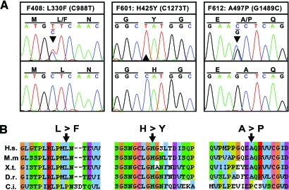
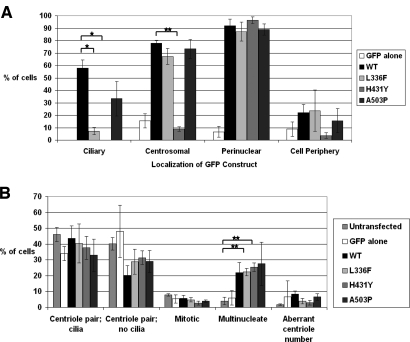
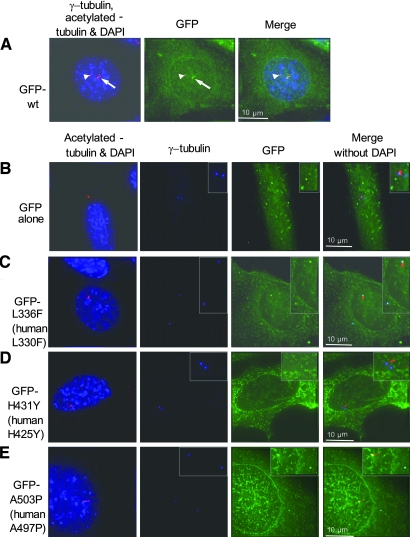
Nephronophthisis, an autosomal recessive kidney disease, is the most frequent genetic cause of chronic renal failure in the first 3 decades of life. Causative mutations in 8 genes (NPHP1-8) have been identified, and homologous mouse models for NPHP2/INVS and NPHP3 have been described. The jck mouse is another model of recessive cystic kidney disease, and this mouse harbors a missense mutation, G448V, in the highly conserved RCC1 domain of Nek8. We hypothesized that mutations in NEK8 might cause nephronophthisis in humans, so we performed mutational analysis in a worldwide cohort of 588 patients. We identified 3 different amino acid changes that were conserved through evolution (L330F, H425Y, and A497P) and that were absent from at least 80 ethnically matched controls. All 3 mutations were within RCC1 domains, and the mutation H425Y was positioned within the same RCC1 repeat as the mouse jck mutation. To test the functional significance of these mutations, we introduced them into full-length mouse Nek8 GFP-tagged cDNA constructs. We transiently overexpressed the constructs in inner medullary collecting duct cells (IMCD-3 cell line) and compared the subcellular localization of mutant Nek8 to wild-type Nek8. All mutant forms of Nek8 showed defects in ciliary localization to varying degrees; the H431Y mutant (human H425Y) was completely absent from cilia and the amount localized to centrosomes was decreased. Overexpression of these mutants did not affect overall ciliogenesis, mitosis, or centriole number. Our genetic and functional data support the assumption that mutations in NEK8 cause nephronophthisis (NPHP9), adding another link between proteins mutated in cystic kidney disease and their localization to cilia and centrosomes.
Figures
Comment in
-
Too much of a good thing: does Nek8 link polycystic kidney disease and nephronophthisis?J Am Soc Nephrol. 2008 Mar;19(3):418-20. doi: 10.1681/ASN.2008010084. Epub 2008 Feb 13. J Am Soc Nephrol. 2008. PMID: 18272836 No abstract available.
References
-
- Watnick T, Germino G: From cilia to cyst. Nat Genet 34: 355–362, 2003 - PubMed
-
- Hildebrandt F, Otto E: Cilia and centrosomes: a unifying pathogenic concept for cystic kidney disease? Nat Rev Genet 6: 928–940, 2005 - PubMed
-
- Hildebrandt F, Otto E, Rensing C, Nothwang HG, Vollmer M, Adolphs J, Hanusch H, Brandis M: A novel gene encoding an SH3 domain protein is mutated in nephronophthisis type 1. Nat Genet 17: 149–153, 1997 - PubMed
-
- Otto E, Hoefele J, Ruf R, Mueller AM, Hiller KS, Wolf MT, Schuermann MJ, Becker A, Birkenhager R, Sudbrak R, Hennies HC, Nurnberg P, Hildebrandt F: A gene mutated in nephronophthisis and retinitis pigmentosa encodes a novel protein, nephroretinin, conserved in evolution. Am J Hum Genet 71: 1161–1167, 2002 - PMC - PubMed
-
- Mollet G, Salomon R, Gribouval O, Silbermann F, Bacq D, Landthaler G, Milford D, Nayir A, Rizzoni G, Antignac C, Saunier S: The gene mutated in juvenile nephronophthisis type 4 encodes a novel protein that interacts with nephrocystin. Nat Genet 32: 300–305, 2002 - PubMed
Publication types
MeSH terms
Substances
Grants and funding
LinkOut - more resources
Full Text Sources
Other Literature Sources
Medical
Molecular Biology Databases
