. 2008 Mar;19(3):503-14.
doi: 10.1681/ASN.2007030300. Epub 2008 Jan 16.

tPA protects renal interstitial fibroblasts and myofibroblasts from apoptosis

Affiliations

tPA protects renal interstitial fibroblasts and myofibroblasts from apoptosis

Kebin Hu et al. J Am Soc Nephrol. 2008 Mar.

Abstract

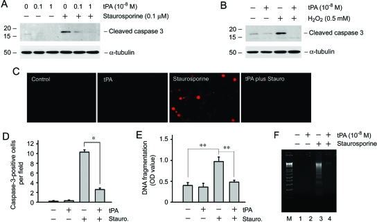
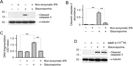
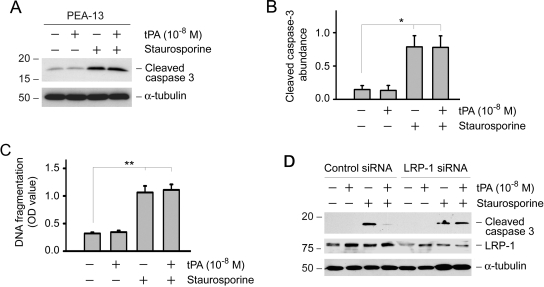
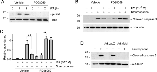
Activation and expansion of interstitial fibroblasts and myofibroblasts play an essential role in the evolution of renal fibrosis. After obstructive injury, mice lacking tissue-type plasminogen activator (tPA) have fewer myofibroblasts and less interstitial fibrosis than wild-type controls. This suggests that tPA controls the size of the fibroblast/myofibroblast population in vivo, and this study sought to determine the underlying mechanism. In vitro, tPA inhibited staurosporine or H(2)O(2)-induced caspase-3 activation, prevented cellular DNA fragmentation, and suppressed the release of cytochrome C from mitochondria into the cytosol in a rat interstitial fibroblast cell line (NRK-49F). tPA also protected TGF-beta1-activated myofibroblasts from apoptosis. This antiapoptotic effect of tPA was independent of its protease activity but required its membrane receptor, the LDL receptor-related protein 1 (LRP-1). Deletion or knockdown of LRP-1 abolished tPA-mediated cell survival, whereas re-introduction of an LRP-1 minigene in a mouse LRP-1-deficient fibroblast cell line (PEA-13) restored the cytoprotective ability of tPA. tPA triggered a cascade of survival signaling involving extracellular signal-regulated kinase 1/2 (Erk1/2), p90RSK, and phosphorylation of Bad. Blockade of Erk1/2 activation abrogated the antiapoptotic effect of tPA, whereas expression of constitutively active MEK1 promoted cell survival similar to tPA. In vivo, compared with wild-type controls, apoptosis of interstitial myofibroblasts was increased in tPA(-/-) mice after obstructive injury, and myofibroblasts were completely depleted 4 wk after relief of the obstruction. Together, these findings illustrate that tPA is a survival factor that prevents apoptosis of renal interstitial fibroblasts and myofibroblasts through an LRP-1-, Erk1/2-, p90RSK-, and Bad-dependent mechanism.


Figures

Figure 1.
Figure 1.
tPA protects renal interstitial fibroblasts from apoptotic death. (A) tPA inhibited staurosporine-induced caspase-3 activation in a dosage-dependent manner. NRK-49F cells were treated with 0.1 μM staurosporine in the absence or presence of various doses of tPA as indicated for 4 h. Cell lysates were immunoblotted with antibodies against cleaved caspase-3 or α-tubulin, respectively. (B) tPA also protected NRK-49F cells from H2O2-triggered caspase-3 activation. NRK-49F cells were incubated with 0.5 mM H2O2 for 16 h. (C and D) Immunofluorescence staining showed that tPA reduced active caspase-3–positive cells after staurosporine treatment. Representative images from various groups (C) as well as quantitative data on active caspase-3–positive cells per ×400 field (D) were presented. *P < 0.05 (n = 5). (E) tPA prevented renal interstitial fibroblasts from DNA fragmentation after staurosporine treatment. DNA fragmentation was assayed at 4 h after various treatments by an ELISA and expressed as OD value at 450 nm. **P < 0.01 (n = 3). (F) tPA prevented NRK-49F cell DNA fragmentation after staurosporine treatment as shown using a DNA laddering assay. M, DNA size marker; 1, control; 2, tPA (10−8 M); 3, staurosporine (0.1 μM); and 4, tPA plus staurosporine.
Figure 2.
Figure 2.
tPA stabilizes mitochondrial membranes and prevents cytochrome C release from mitochondria. (A) Mitochondria and cytosol of NRK-49F cells were separated after staurosporine treatment in the absence or presence of tPA. Cytosolic proteins were subjected to Western blot analysis for cytochrome C. (B) Quantitative determination of the relative abundance of cytochrome C release among different groups. Data are means ± SEM of three experiments. **P < 0.01. (C) Immunofluorescence staining for cytochrome C (green) in NRK-49F cells after various treatments as indicated. DAPI (blue) was used to visualize the nuclei.
Figure 3.
Figure 3.
tPA protects renal interstitial myofibroblasts from apoptosis in vitro. NRK-49F cells were preincubated with 0.5 ng/ml TGF-β1 for 24 h to induce the activated myofibroblast phenotype, as confirmed by positive staining for α-SMA. Cells were then treated with staurosporine (0.1 μM) and/or tPA as indicated for 4 h. (A) Double immunofluorescence staining for α-SMA (green) and active caspase-3 (red). Nuclei were stained with DAPI (blue). (B and C) Western blot analysis demonstrated the presence of active, cleaved caspase-3. A representative picture (B) and quantitative determination (C) of relative cleaved caspase-3 levels are presented. *P < 0.05 (n = 3).
Figure 4.
Figure 4.
Antiapoptotic effect of tPA is independent of its protease activity. (A) NRK-49F cells were incubated with staurosporine (0.1 μM) and/or nonenzymatic tPA (10−8 M) for 4 h. Cell lysates were immunoblotted with antibodies against active, cleaved caspase-3 and α-tubulin, respectively. (B) Quantitative determination of relative abundance of cleaved caspase-3. Data are means ± SEM of three experiments. **P < 0.01. (C) Nonenzymatic tPA prevented DNA fragmentation in NRK-49F cells after staurosporine treatment for 4 h. DNA fragmentation was assayed by an ELISA and expressed as OD value at 450 nm. **P < 0.01 (n = 3). (D) MMP-9 did not affect fibroblast apoptosis after staurosporine treatment. NRK-49F cells were treated with staurosporine (0.1 μM) and/or MMP-9 (10−8 M) for 4 h. Cell lysates were immunoblotted with antibodies against cleaved caspase-3 and α-tubulin, respectively.
Figure 5.
Figure 5.
Antiapoptotic effect of tPA is mediated by its cell membrane receptor LRP-1. (A) Knockout of LRP-1 abolished the prosurvival action of tPA. LRP-1–deficient mouse embryonic fibroblast cells (PEA-13) were incubated with staurosporine (0.1 μM) and/or tPA (10−8 M) for 6 h. Cell lysates were immunoblotted with antibodies against cleaved caspase-3 and α-tubulin, respectively. (B) Quantitative determination of relative abundance of cleaved caspase-3. Data are means ± SEM of three experiments. *P < 0.05. (C) DNA fragmentation was assayed by an ELISA and expressed as OD 450 nm. **P < 0.01 (n = 3). (D) Knockdown of LRP-1 in NRK-49F cells by siRNA inhibition abolished the cytoprotective effect of tPA. NRK-49F cells were transfected with either control siRNA or LRP-1–specific siRNA, followed by treatment with staurosporine and/or tPA as indicated. Cell lysates were immunoblotted with antibodies against cleaved caspase-3, LRP-1, and α-tubulin.
Figure 6.
Figure 6.
mini-LRP-1 gene restores the cytoprotective effect of tPA in LRP−/− fibroblasts. (A) Graphic illustration of the structure of full-length and mini-LRP-1. mLRP2 consists of the ligand-binding domain II, which binds to tPA, and the entire 85-kD β unit of LRP-1. (B) Overexpression of pHA-mLRP2 into LRP-1−/− fibroblast (PEA-13) restored the antiapoptotic effect of tPA. (C) Quantitative illustration of relative abundance of cleaved caspase 3. **P < 0.01 (n = 3).
Figure 7.
Figure 7.
tPA activates cell survival signaling via sequential phosphorylation of Erk1/2, p90RSK, and Bad. NRK-49F cells were incubated with 10−8 M tPA for various periods of time as indicated and then subjected to Western blotting for phospho-Erk1/2 (A), phospho-p90RSK (B), and phospho-Bad (C). The samples were also probed with antibodies against total Erk1/2, p90RSK1/2/3, and Bad, respectively. (D) Schematic illustration of the cytoprotective effects of tPA in interstitial fibroblast. After binding to its membrane receptor LRP-1, tPA induces its tyrosine phosphorylation, which in turn stimulates Erk1/2 and p90RSK phosphorylation, leading to Bad phosphorylation. Phosphorylation of Bad suppresses cytochrome C release from mitochondria and protects the cells from apoptosis. *Tyrosine or serine/threonine phosphorylation.
Figure 8.
Figure 8.
Erk1/2 activation is indispensable for mediating interstitial fibroblast survival induced by tPA. (A) Pretreatment with the MEK1 inhibitor PD98059 (20 μM) for 0.5 h abrogated the Bad phosphorylation induced by tPA. (B) PD98059 also abolished the tPA-mediated inhibition of caspase-3 activation in NRK-49F cells. (C) Quantitative determination of relative abundance of cleaved caspase-3. Data are means ± SEM of three experiments. **P < 0.01. (D) Overexpression of constitutively active MEK1 by an adenoviral vector (Ad.MEK1) was sufficient to prevent caspase-3 activation after staurosporine treatment. NRK-49F cells were infected with adenovirus for 16 h, followed by incubation with staurosporine (0.1 μM) for 4 h. An adenoviral vector containing the β-galactosidase gene (Ad.LacZ) was used as a control.
Figure 9.
Figure 9.
Increased apoptosis of interstitial myofibroblasts in mice lacking tPA after obstructive injury. (A) Western blot analyses show a reduced α-SMA expression in tPA−/− mice after obstructive injury. Kidney homogenates from both tPA+/+ and tPA−/− mice at 7 d after UUO were assayed for α-SMA expression. Contralateral intact kidneys (CLK) served as controls. (B) Quantitative determination of relative abundance of α-SMA protein among various groups. Data are means ± SEM of four to five animals per group. *P < 0.05. (C) Representative micrographs show immunohistochemical staining for cleaved caspase-3 (red, left). (Middle, inset) Enlarged image of the cleaved caspase-3–positive cell. (Right) Double immunofluorescence staining confirmed the co-localization of α-SMA (green) and cleaved caspase-3 (red). Paraffin-embedded kidney sections were prepared from tPA+/+ or tPA−/− mice at day 7 after UUO. Arrowheads indicate the cells with positive staining in the obstructed kidneys. (D) Interstitial cells undergoing apoptosis (cleaved caspase-3 positive) were calculated and expressed as the percentage in total interstitial cell population. Data were obtained from five animals per group. **P < 0.01 (n = 5). (E) Experimental design for reversible UUO model. (F) Western blot analyses show α-SMA expression at 4 wk after relief of obstruction in the reversible UUO model. Complete loss of α-SMA expression was observed only in tPA−/− mice but not in tPA+/+ controls. Numbers 1 through 5 denote each individual animal. (G) Quantitative determination of α-SMA expression at 4 wk after relief of obstruction in reversible UUO model. *P < 0.05 (n = 5).

References

    1. Liu Y: Renal fibrosis: New insights into the pathogenesis and therapeutics. Kidney Int 69: 213–217, 2006 - PubMed
    1. Eddy AA: Progression in chronic kidney disease. Adv Chronic Kidney Dis 12: 353–365, 2005 - PubMed
    1. Strutz F, Zeisberg M: Renal fibroblasts and myofibroblasts in chronic kidney disease. J Am Soc Nephrol 17: 2992–2998, 2006 - PubMed
    1. Qi W, Chen X, Poronnik P, Pollock CA: The renal cortical fibroblast in renal tubulointerstitial fibrosis. Int J Biochem Cell Biol 38: 1–5, 2006 - PubMed
    1. Neilson EG: Mechanisms of disease: Fibroblasts—A new look at an old problem. Nat Clin Pract Nephrol 2: 101–108, 2006 - PubMed

Publication types

MeSH terms

Substances