. 2008 Apr 1;111(7):3343-54.
doi: 10.1182/blood-2007-10-115758. Epub 2008 Jan 16.

HIF-1alpha regulates epithelial inflammation by cell autonomous NFkappaB activation and paracrine stromal remodeling

Affiliations

HIF-1alpha regulates epithelial inflammation by cell autonomous NFkappaB activation and paracrine stromal remodeling

Marzia Scortegagna et al. Blood. .

Abstract

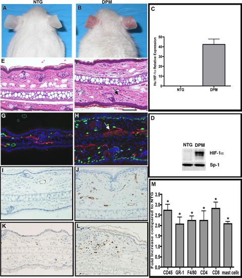
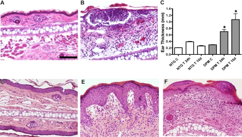
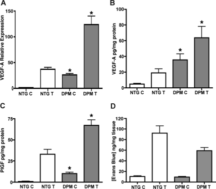
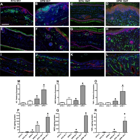
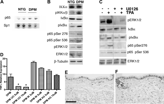
Hypoxia inducible factor-1 (HIF-1) is a master regulatory transcription factor controlling multiple cell-autonomous and non-cell-autonomous processes, such as metabolism, angiogenesis, matrix invasion, and cancer metastasis. Here we used a new line of transgenic mice with constitutive gain of HIF-1 function in basal keratinocytes and demonstrated a signaling pathway from HIF-1 to nuclear factor kappa B (NFkappaB) activation to enhanced epithelial chemokine and cytokine elaboration. This pathway was responsible for a phenotypically silent accumulation of stromal inflammatory cells and a marked inflammatory hypersensitivity to a single 12-O-tetradecanoylphorbol-13-acetate (TPA) challenge. HIF-1-induced NFkappaB activation was composed of 2 elements, IkappaB hyperphosphorylation and phosphorylation of Ser276 on p65, enhancing p65 nuclear localization and transcriptional activity, respectively. NFkappaB transcriptional targets macrophage inflammatory protein-2 (MIP-2/CXCL2/3), keratinocyte chemokine (KC/CXCL1), and tumor necrosis factor [alfa] (TNFalpha) were constitutively up-regulated and further increased after TPA challenge both in cultured keratinocytes and in transgenic mice. Whole animal KC, MIP-2, or TNFalpha immunodepletion each abrogated TPA-induced inflammation, whereas blockade of either VEGF or placenta growth factor (PlGF) signaling did not affect transgenic inflammatory hyper-responsiveness. Thus, epithelial HIF-1 gain of function remodels the local environment by cell-autonomous NFkappaB-mediated chemokine and cytokine secretion, which may be another mechanism by which HIF-1 facilitates either inflammatory diseases or malignant progression.


Figures

Figure 1
Figure 1
“Inflammatory priming” by epithelial mediated stromal inflammatory cell recruitment in K14-HIF-1αDPM transgenic mice. Redness and prominent vasculature of ear skin in transgenic mice (DPM) (B), which are not evident in nontransgenic mice (NTG) (A). Human HIF-1α mRNA (C) and total (human and potentially mouse) HIF-1α protein (D) are detectable only in transgenic ears. Ear histology reveals increased dermal cellularity in transgenic mice (F, arrows), compared with nontransgenic mice (E). Differential increase in subepidermal microvessels, revealed by MECA-32 immunohistochemistry (green), and lymphatic dilatation, LYVE-1 antibody (red, see white arrow) in DPM transgenic mice (H) versus nontransgenic controls (G). Endothelial activation evidenced by ICAM-1 immunohistochemistry, in transgenic (J), versus nontransgenic (I) ears. Increased number of CD45.1 positive inflammatory cells in transgenic (L) versus nontransgenic (K) ears. A 2- to 3-fold increase of neutrophils, mast cells, macrophages, and lymphocytes, determined by differential immunohistochemical analysis of inflammatory markers as indicated in panel M. Bar in panel F represents 100 μm.
Figure 2
Figure 2
Transgenic mice exhibit enhanced and prolonged response to an inflammatory challenge. Intraepidermal neutrophilic abscesses 6 hours after TPA challenge in transgenic mice (B) compared with low-level dermal inflammatory cell infiltrate in nontransgenic controls (A). Persistent ear swelling 10 days after TPA challenge evidenced by caliper ear thickness in transgenic (DPM) versus control (NTG) ears (C). Histologic resolution of inflammation in nontransgenic ears 10 days after TPA challenge (D). In contrast, transgenic ears (E,F) evidence psoriatic-type changes of increased epidermal thickness, epidermal rete ridge dermal projections, subepidermal edema, and foci of parakeratosis (F). Bar in panel A represents 100 μm.
Figure 3
Figure 3
Increased VEGF-A and PlGF protein levels without microvascular leakage in transgenic mice. Real-time TaqMan RT-PCR analysis of VEGF-A expression from total RNA isolated from ears of K14-HIF-1αDPM transgenic mice (DPM) and nontransgenic controls (NTG), using histone 3.3A as a reference (A), at baseline “C” and after 6 hours of TPA treatment “T.” VEGF-A (B) and PlGF (C) ELISA assays reveal significantly higher protein expression in untreated and TPA treated transgenic ears compared with nontransgenic controls. Lack of differential microvascular leakage, evidenced by ear Evans blue dye content, in transgenic vs nontransgenic mice (D). Error bars represent mean plus or minus SEM of 4 to 6 mice analyzed per group (*P < .05, Student t test).
Figure 4
Figure 4
K14-HIF-1αDPM mice exhibit epidermal and dermal inflammatory infiltrates accompanied by hyperplastic cutaneous blood vessels and lymphovascular dilation. Increase of GR1+ neutrophils in green (A-D) and F4/80+ macrophages in green (E-H) 6 hours (B,F) and 10 days after TPA challenge (D,H) in transgenic ears (DPM) compared with nontransgenic littermates (NTG) (A, E, C, and G, respectively). Counterstaining with a keratin-14 antibody (red) highlights the differential persistent increase in epidermal thickness in transgenic mice. Progressive increase in microvascular density (MECA32, green) along with lymphovascular dilatation (LYVE-1, red) is observed between the 6-hour and 10-day after TPA time intervals in transgenic ears (J,L) compared with nontransgenic littermates (I,K). Bar in panel A represents 100 μm. Real-time TaqMan RT-PCR analysis of keratinocyte chemokine (KC) (M), macrophage inflammatory protein-2 (MIP-2) (N), and TNFα (O) expression from total RNA isolated from ears of transgenic and nontransgenic controls, using histone 3.3A as a reference, at baseline and 1 hour after TPA treatment. KC (P), MIP-2 (Q), and TNFα. (R) ELISA assays reveal significantly higher protein expression in the inflamed ears of transgenic mice after 3 hours and 10 days after a single TPA application. Error bars represent mean plus or minus SEM of 4 to 6 mice analyzed per group (*P < .05).
Figure 5
Figure 5
Epithelial inflammatory chemokine elaboration requires NFκB signaling. Elevation of KC, MIP-2, and VEGF-A protein expression at baseline “C” and 3 hours after TPA treatment “T” in supernatants conditioned by primary transgenic keratinocytes (A, B, and C, respectively) demonstrate the cell autonomous activity of epithelial HIF-1α gain of function. (D) Increased NFκB transcriptional activity in K14-HIF-1αDPM keratinocytes. Nontransgenic (NTG) and transgenic (DPM) keratinocytes were transiently transfected with a NFκB reporter construct. After 48 hours, cells were treated with DMSO or TPA for 3 hours, harvested, and assayed for luciferase activity. Values are expressed as relative light units per microgram total protein (D). IκBαSer32/34A (IκBα super-repressor, IκBαSR) NFκB transcriptional blockade markedly diminishes keratinocyte chemokine expression (KC, MIP-2, and TNF-α, panels E-G, respectively) but does not affect the HIF-1α target gene (VEGF-A, PlGF, and Glut-1, panels H-J, respectively) expression. RT-PCR analysis using total RNA extracted from a control adenovirus (A-cytomegalovirus) or IκBαSR adenoviral-transduced primary keratinocytes at baseline and 3 hours after TPA treatment. Error bars represent mean plus or minus SEM. Results are representative of 3 independent experiments (*P < .05, t test).
Figure 6
Figure 6
NFκB activation in K14-HIF-1αDPM keratinocytes is dependent on increased Erk1/2 phosphorylation. Increased nuclear translocation of the NFκB subunit p65 in transgenic (DPM) versus nontransgenic keratinocytes (NTG), normalized to Sp1 (A). Nuclear extracts were analyzed by Western blots. Western blot of lysates from newborn transgenic (DPM) keratinocytes indicate a 2- to 3-fold increase of phosphorylated IKKα, IκBα, p65 phosphorylation at serines 276 and 536, and phospho-ERK1/2, compared with nontransgenic (NTG) keratinocytes; β-tubulin is a loading control (B). Transgenic keratinocytes were pretreated with DMSO (−) or the MEK1/2 inhibitor U0126 (+) for 24 hours and analyzed at baseline (−) or 10 minutes after TPA treatment (+). Western blots of lysates from newborn transgenic keratinocytes show inhibition of phospho-ERK1/2 and p65 Ser276 phosphorylation by U0126 both at baseline and after TPA treatment (C). In contrast, phospho-IκBα, phospho-p65 Ser536, and the level of IκBα protein remain unaffected by ERK1/2 inhibition (C); total ERK1/2 is used as a loading control. The separating line in panel C represents one lane deleted from the same gel. Real-time RT-PCR analysis of TNFα expression from total RNA extracted from transgenic (DPM) primary keratinocytes shows selective inhibition of TNFα expression 24 hours after U0126 (U0) treatment in contrast to PI3 kinase pathway wortmannin (W), or EGFR AG1478 (AG) inhibition, both of which failed to alter transgenic keratinocyte TNFα expression (D). Bars represent mean plus or minus SEM. Results are representative of 3 independent experiments (*P < .05, t test). (E-F) Increased ERK1/2 phosphorylation is also evident in immunohistochemical analysis of transgenic compared with nontransgenic ears (E,F). Bar in panel E represents 100 μm.
Figure 7
Figure 7
Immunodepletion of candidate chemokines, cytokines, and growth factors demonstrate selective dependence of HIF-1α–mediated inflammatory hyper-responsiveness on NFκB targets. Transgenic mice were pretreated with neutralizing antibodies targeting either KC (A), a KC/MIP-2 cocktail (B), TNFα (C), or a VEGFR1/VEGFR2 cocktail (D). Twenty-four hours later, a single dose of TPA (2.5 μg) was applied to the ear followed by tissue harvest 6 hours later and hematoxylin and eosin histostaining. Immunodepletion with either KC or TNFα antibodies markedly decreased, whereas the KC/MIP-2 cocktail abrogated, HIF-1α–mediated inflammatory hyper-responsiveness. In marked contrast, transgenic ears were resistant to VEGFR1/VEGFR2 immunodepletion. Immunofluorescence for GR1 expression confirms that KC/MIP-2 depletion abrogates neutrophil recruitment to TPA-challenged transgenic ears (E, GR1, green; K14, red). In contrast, immunodepleted, TPA-challenged transgenic ears retain elevated subepidermal microvascular density and lymphatic dilatation (F, MECA32, green; LYVE-1, red). Decreased immunofluorescence for ICAM1 on TPA-challenged transgenic ears pretreated with VEGFR1/2 neutralizing antibodies (H, ICAM-1, green; MECA32, red) versus nonpretreated and TPA-challenged transgenic ears (G, ICAM-1, green; MECA32, red) confirms the effectiveness of VEGFR1/2 antibody treatment. Bar in panel A represents 100 μm.

References

    1. Karin M, Lawrence T, Nizet V. Innate immunity gone awry: linking microbial infections to chronic inflammation and cancer. Cell. 2006;124:823–835. - PubMed
    1. de Visser KE, Eichten A, Coussens LM. Paradoxical roles of the immune system during cancer development. Nat Rev Cancer. 2006;6:24–37. - PubMed
    1. Zenz R, Eferl R, Kenner L, et al. Psoriasis-like skin disease and arthritis caused by inducible epidermal deletion of Jun proteins. Nature. 2005;437:369–375. - PubMed
    1. Sano S, Chan KS, Kira M, et al. Signal transducer and activator of transcription 3 is a key regulator of keratinocyte survival and proliferation following UV irradiation. Cancer Res. 2005;65:5720–5729. - PubMed
    1. Semenza G. Signal transduction to hypoxia-inducible factor 1. Biochem Pharmacol. 2002;64:993–998. - PubMed

Publication types

MeSH terms