. 2008 Jan 22;105(3):859-64.
doi: 10.1073/pnas.0709813105. Epub 2008 Jan 16.

The intersection between cell wall disassembly, ripening, and fruit susceptibility to Botrytis cinerea

Affiliations

The intersection between cell wall disassembly, ripening, and fruit susceptibility to Botrytis cinerea

D Cantu et al. Proc Natl Acad Sci U S A. .

Abstract

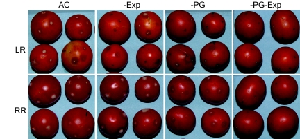
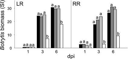
Fruit ripening is characterized by processes that modify texture and flavor but also by a dramatic increase in susceptibility to necrotrophic pathogens, such as Botrytis cinerea. Disassembly of the major structural polysaccharides of the cell wall (CW) is a significant process associated with ripening and contributes to fruit softening. In tomato, polygalacturonase (PG) and expansin (Exp) are among the CW proteins that cooperatively participate in ripening-associated CW disassembly. To determine whether endogenous CW disassembly influences the ripening-regulated increase in necrotropic pathogen susceptibility, B. cinerea susceptibility was assessed in transgenic fruit with suppressed polygalacturonase (LePG) and expansin (LeExp1) expression. Suppression of either LePG or LeExp1 alone did not reduce susceptibility but simultaneous suppression of both dramatically reduced the susceptibility of ripening fruit to B. cinerea, as measured by fungal biomass accumulation and by macerating lesion development. These results demonstrate that altering endogenous plant CW disassembly during ripening influences the course of infection by B. cinerea, perhaps by changing the structure or the accessibility of CW substrates to pathogen CW-degrading enzymes. Recognition of the role of ripening-associated CW metabolism in postharvest pathogen susceptibility may be useful in the design and development of strategies to limit pathogen losses during fruit storage, handling, and distribution.


Conflict of interest statement

The authors declare no conflict of interest.

Figures

Fig. 1.
Fig. 1.
LePG and LeExp1 expression influences the softening of ripening Ailsa Craig (AC) tomato fruit. (A) Western blots demonstrate that proteins recognized by antibodies to tomato PG and Exp are present in red ripe (RR) AC fruit, that PG is absent in −PG fruit, that Exp is absent in −Exp fruit, and that both proteins are missing from the −PG−Exp fruit. (B) Measuring the force to compress each fruit 2 mm shows that decreasing PG and Exp in fruit significantly reduces softening at all ripening stages. Firmness was measured at the mature green (MG) stage and in subsequent ripening stages [breaker (BR), light red (LR), and RR]. Letters correspond to significant differences between genotypes at each ripening stage; P < 0.05.
Fig. 2.
Fig. 2.
Reduction of PG and Exp substantially decreases susceptibility to B. cinerea. LR and RR fruit were inoculated with B. cinerea spores at three of four puncture sites on each fruit (the fourth site was mock-inoculated with water), and disease symptoms were assessed at 1, 2, and 3 dpi. Fruit showed disease symptoms when an expanding macerating (water-soaked) lesion developed at any inoculated site, and the percentage of the fruit showing any symptoms of disease is shown. Approximately 50 fruit from each genotype were analyzed and the experiment was done at least twice.
Fig. 3.
Fig. 3.
Reduction of PG and Exp substantially decreases gray mold symptoms in inoculated LR or RR fruit. The image is of fruit at 3 dpi.
Fig. 4.
Fig. 4.
Reduction of PG and Exp decreases pathogen growth on fruit and the CW composition from infected tissue demonstrates that fungal biomass accumulation is reduced on −PG−Exp fruit. (A) Infected tissues were excised 36 and 72 h after inoculation (hpi), and the assessment of B. cinerea biomass in homogenates was based on signal intensity (SI) using the monoclonal antibody BC12.CA4. Letters correspond to significant differences between genotypes at each hpi; P < 0.001. (B) AIRs were prepared from infected tissues and analyzed colorimetrically for UA and NS contents, expressed as the UA/NS ratios. Block letters correspond to significant differences between genotypes at each hpi; italic letters correspond to significant differences between hpi for each genotype; P < 0.001. (C) AIR from RR fruit at 36 and 72 hpi were hydrolyzed and derivatized to determine the NS by GC-MS. GC-MS detector output is shown. Elution times of the alditol acetate derivative peaks are shown. Ara, arabinose; Xyl, xylose; Man, mannose; Gal, galactose; Glc, glucose; Ino, myo-inositol internal standard. The relative abundance of each sugar is expressed as the total ion count. (I) AC, 36 hpi. (II) AC, 72 hpi. (III) −PG−Exp, 36 hpi. (IV) −PG−Exp, 72 hpi. The elevated Man and Glc indicate the increased B. cinerea CW contents in AC fruit AIR. Identification of bars is as defined in Fig. 1B.
Fig. 5.
Fig. 5.
Pathogen growth is reduced in liquid cultures containing −PG−Exp CWs compared with cultures supplied with AC CWs. Mycelia were collected 1, 3, and 6 dpi from cultures containing AIR from uninfected LR and RR fruit of the four genotypes. Fungal biomass was measured as described in the legend of Fig. 4A. Identification of bars is as defined in Fig. 1B. Letters correspond to significant differences between genotypes at each dpi; P < 0.001.
Fig. 6.
Fig. 6.
Reduction of PG and Exp in ripening fruit decreases the solubilization and depolymerization of pectin polysaccharides. Pectins were extracted sequentially from LR and RR AIR from the four genotypes by using water, CDTA, and Na2CO3. (A) Histograms of the distribution of UAs in the pectin extracts. (B) Images show the size exclusion chromatographic fractionation of the water-soluble pectins (WSF) from AC and −PG−Exp fruit at the LR and RR ripening stages. The data show that neither the normal ripening-associated increase in fruit WSF content nor the decrease in polymer size, both observed in AC fruit, occurs in −PG−Exp fruit.
Fig. 7.
Fig. 7.
The CW swells as AC tomato fruit ripen but not as −PG−Exp fruit ripen. (A) Electron micrographs show the increased thickness of walls (CW) from RR fruit pericarp cells and increased spacing of the wall's electron-dense fibrillar material in AC compared with −PG−Exp fruit. (Scale bars, 1 μm.) (B) The thickness of CW from 30 images as in A show that walls from RR AC (black bar) fruit are thicker than walls from RR −PG−Exp (white bar) fruit. Letters indicate significant differences between genotypes; P < 0.01. (C) By comparing the depth of the settled aqueous suspensions of AIR from LR and RR AC fruit, a typical ripening-associated AIR swelling is observed. No such change is seen when LR and RR AIR samples from −PG−Exp fruit are allowed to settle. (Scale bars, 1 cm.)

References

    1. Vorwerk S, Somerville S, Somerville C. Trends Plant Sci. 2004;9:203–209. - PubMed
    1. Hückelhoven R, editor. Annu Rev Phytopath. 2007;45:101–127. - PubMed
    1. Collmer A, Keen NT. Annu Rev Phytopath. 1986;24:383–409.
    1. Brummell DA, Howie WJ, Ma C, Dunsmuir P. Postharvest Biol Tech. 2002;25:209–220.
    1. ten Have A, Mulder W, Visser J, van Kan JAL. Mol Plant–Microbe Interact. 1998;11:1009–1016. - PubMed

Publication types

MeSH terms

LinkOut - more resources