. 2008 Jan 22;105(3):985-90.
doi: 10.1073/pnas.0711281105. Epub 2008 Jan 16.

PPARalpha agonist fenofibrate suppresses tumor growth through direct and indirect angiogenesis inhibition

Affiliations

PPARalpha agonist fenofibrate suppresses tumor growth through direct and indirect angiogenesis inhibition

Dipak Panigrahy et al. Proc Natl Acad Sci U S A. .

Abstract

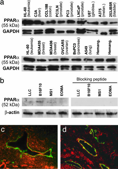
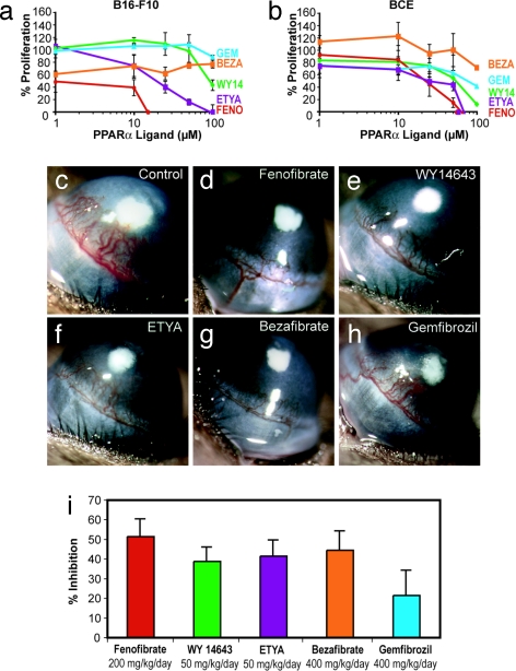
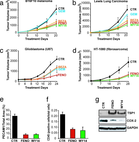
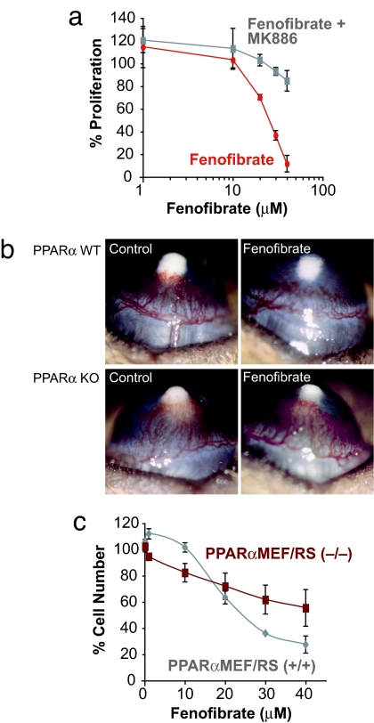
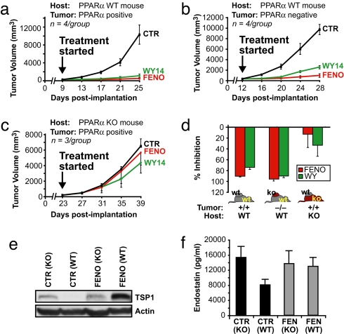
Angiogenesis and inflammation are central processes through which the tumor microenvironment influences tumor growth. We have demonstrated recently that peroxisome proliferator-activated receptor (PPAR)alpha deficiency in the host leads to overt inflammation that suppresses angiogenesis via excess production of thrombospondin (TSP)-1 and prevents tumor growth. Hence, we speculated that pharmacologic activation of PPARalpha would promote tumor growth. Surprisingly, the PPARalpha agonist fenofibrate potently suppressed primary tumor growth in mice. This effect was not mediated by cancer-cell-autonomous antiproliferative mechanisms but by the inhibition of angiogenesis and inflammation in the host tissue. Although PPARalpha-deficient tumors were still susceptible to fenofibrate, absence of PPARalpha in the host animal abrogated the potent antitumor effect of fenofibrate. In addition, fenofibrate suppressed endothelial cell proliferation and VEGF production, increased TSP-1 and endostatin, and inhibited corneal neovascularization. Thus, both genetic abrogation of PPARalpha as well as its activation by ligands cause tumor suppression via overlapping antiangiogenic pathways. These findings reveal the potential utility of the well tolerated PPARalpha agonists beyond their use as lipid-lowering drugs in anticancer therapy. Our results provide a mechanistic rationale for evaluating the clinical benefits of PPARalpha agonists in cancer treatment, alone and in combination with other therapies.


Conflict of interest statement

The authors declare no conflict of interest.

Figures

Fig. 1.
Fig. 1.
PPARα is expressed in tumor cells and endothelium of neoplastic tissues. (a) Western blot analysis of PPARα expression in cultured human tumor cells and hemangioma specimens. Nuclear extract from leukemia cells (HL-60) was used as a control. (b) Western blot analysis of PPARα expression in cultured mouse tumor cells. The specificity of protein expression was confirmed by abrogation by a PPARα-blocking peptide. GAPDH and β-actin levels were measured to demonstrate equal loading of protein in each lane. (c and d) Immunofluorescent double staining for CD31 and PPARα demonstrates PPARα expression in endothelium of human pancreatic cancer (BxPC3) in SCID mice (c) and in patient prostate cancer tissue specimens (d). CD31-stained endothelial cells are shown in green, PPARα-positive cells are red, and colocalization of the two colors are yellow. Colocalization of red and green fluorescence (yellow) indicates PPARα expression in blood vessels.
Fig. 2.
Fig. 2.
PPARα ligands have direct antitumor and antiendothelial effects in vitro and in vivo. (a) Percentage of proliferation of tumor cells (B16-F10 melanoma) is determined by comparing cells grown in media plus 10% FBS, and the PPARα ligands, to starved cells. FENO, fenofibrate; WY14, WY14643; BEZA, bezafibrate; GEM, gemfibrozil. (b) Percentage of proliferation of BCE cells is determined by comparing cells exposed to an angiogenic stimulus (FGF2) with those exposed to FGF2 and PPARα ligands (fenofibrate, WY14643, gemfibrozil, ETYA, and bezafibrate) relative to unstimulated cells. (c) FGF2-induced neovascularization in control cornea on day 6 in a mouse receiving vehicle. (d–h) Systemic treatment with fenofibrate at 200 mg/kg per day (d), WY14643 at 50 mg/kg per day (e), ETYA at 50 mg/kg per day (f), bezafibrate at 400 mg/kg per day (g), or gemfibrozil at 400 mg/kg per day (h). (i) Area of inhibition (percentage) by administration of various PPARα ligands: fenofibrate (200 mg/kg per day), 52% inhibition; WY14643 (50 mg/kg per day), 39% inhibition; ETYA (50 mg/kg per day), 42% inhibition; bezafibrate (400 mg/kg per day), 44% inhibition; and gemfibrozil (400 mg/kg per day), 22% inhibition. Inhibition was determined on day 6 by the following formula: pellet distance × 0.2π × neovessel length × clock hours of neovessels. (n = 6 eyes per group; the experiment was performed three times.)
Fig. 3.
Fig. 3.
Systemic therapy with PPARα ligands inhibits primary tumor growth. When tumors reached 100 mm3 in size, PPARα ligand treatment was initiated (day 0). On the last day of treatment, the statistical difference between control and treated group was determined by Student's t test. The most potent antitumor activity was obtained by fenofibrate and WY14643 at the following doses: fenofibrate, 200 mg/kg per day; WY14643, 50 mg/kg per day; bezafibrate, 200 mg/kg per day; and gemfibrozil, 200 mg/kg per day. (a) B16-F10 melanoma (P < 0.001). (b) LLC (P < 0.001). (c) Glioblastoma (U87) (P < 0.005). (d) Fibrosarcoma (HT-1080) (P < 0.0001). (e) Vessel density in fenofibrate-, WY14643-, and vehicle-treated B16-F10 tumors, as defined by the percentage of vessel area = PECAM1-positive area/tumor area in each field. (f) Leukocyte counts per total number of cells per field in fenofibrate-treated and WY14643-treated and vehicle-treated B16-F10 tumors, as determined by CD45 staining. (g) Western blot analysis of TSP-1 and COX-2 proteins in tumor lysates of fenofibrate-, WY14643-, and vehicle-treated B16-F10 melanomas on day 20.
Fig. 4.
Fig. 4.
PPARα ligands have PPARα-dependent and -independent effects. (a) Effect of fenofibrate with or without MK886 treatment on the percentage of proliferation on endothelial cells. (b) Corneal neovascularization (80 ng of FGF2 pellets) in fenofibrate- and vehicle-treated PPARα WT and PPARα KO mice. (c) Effect of fenofibrate treatment on proliferation of PPARα-positive tumor PPARα+/+MEF/RS and PPARα-negative tumor PPARα−/−MEF/RS on day 3.
Fig. 5.
Fig. 5.
The antitumor activity of PPARα ligands is host PPARα receptor-dependent. After PPARα-positive (PPARα+/+MEF/RS) or PPARα-negative (PPARα−/−MEF/RS) tumors were 100 mm3 in size, PPARα ligand treatment was initiated (day 0). The doses were as follows: fenofibrate, 200 mg/kg per day; and WY14643, 50 mg/kg per day. (a) The effect of systemic therapy of PPARα ligands on PPARα-positive tumors (PPARα+/+MEF/RS) in PPARα WT mice (90–95% inhibition). (b) The effect of systemic therapy on PPARα-negative tumors (PPARα−/−MEF/RS) in PPARα WT (75–90% inhibition). (c) The effect of systemic therapy of PPARα-positive tumors (PPARα+/+MEF/RS) in PPARα (KO) mice (13–33% inhibition). (d) The columns summarize the effects of fenofibrate and WY14643 in host or tumor cells. (e) Western blot analysis of TSP-1 in plasma from fenofibrate and vehicle treated PPARα WT and PPARα KO mice. (f) Endostatin levels in plasma from fenofibrate- and vehicle-treated PPARα WT and PPARα KO mice.

References

    1. Lefebvre P, Chinetti G, Fruchart JC, Staels B. Sorting out the roles of PPARα in energy metabolism and vascular homeostasis. J Clin Invest. 2006;116:571–580. - PMC - PubMed
    1. Devchand PR, et al. The PPARα-leukotriene B4 pathway to inflammation control. Nature. 1996;384:39–43. - PubMed
    1. Peters JM, Cattley RC, Gonzalez FJ. Role of PPARα in the mechanism of action of the nongenotoxic carcinogen and peroxisome proliferator Wy 14,643. Carcinogenesis. 1997;18:2029–2033. - PubMed
    1. Michalik L, Desvergne B, Wahli W. Peroxisome-proliferator-activated receptors and cancers: complex stories. Nat Rev Cancer. 2004;4:61–70. - PubMed
    1. Collett GP, et al. Peroxisome proliferator-activated receptorα is an androgen-responsive gene in human prostate and is highly expressed in prostatic adenocarcinoma. Clin Cancer Res. 2000;6:3241–3248. - PubMed

Publication types

MeSH terms

LinkOut - more resources