. 2008 Jan 22;105(3):889-94.
doi: 10.1073/pnas.0710728105. Epub 2008 Jan 16.

Micromechanical properties of keratin intermediate filament networks

Affiliations

Micromechanical properties of keratin intermediate filament networks

Sivaraj Sivaramakrishnan et al. Proc Natl Acad Sci U S A. .

Abstract

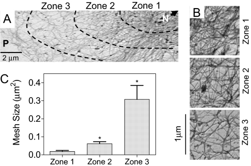
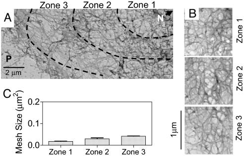
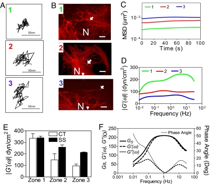
Keratin intermediate filaments (KIFs) form cytoskeletal KIF networks that are essential for the structural integrity of epithelial cells. However, the mechanical properties of the in situ network have not been defined. Particle-tracking microrheology (PTM) was used to obtain the micromechanical properties of the KIF network in alveolar epithelial cells (AECs), independent of other cytoskeletal components, such as microtubules and microfilaments. The storage modulus (G') at 1 Hz of the KIF network decreases from the perinuclear region (335 dyn/cm(2)) to the cell periphery (95 dyn/cm(2)), yielding a mean value of 210 dyn/cm(2). These changes in G' are inversely proportional to the mesh size of the network, which increases approximately 10-fold from the perinuclear region (0.02 microm(2)) to the cell periphery (0.3 microm(2)). Shear stress (15 dyn/cm(2) for 4 h) applied across the surface of AECs induces a more uniform distribution of KIF, with the mesh size of the network ranging from 0.02 microm(2) near the nucleus to only 0.04 microm(2) at the cell periphery. This amounts to a 40% increase in the mean G'. The storage modulus of the KIF network in the perinuclear region accurately predicts the shear-induced deflection of the cell nucleus to be 0.87 +/- 0.03 microm. The high storage modulus of the KIF network, coupled with its solid-like rheological behavior, supports the role of KIF as an intracellular structural scaffold that helps epithelial cells to withstand external mechanical forces.


Conflict of interest statement

The authors declare no conflict of interest statement.

Figures

Fig. 1.
Fig. 1.
Mesh size distribution of the KIF network in control primary rat AECs. (A) Representative transmission electron micrograph of primary rat AECs, showing in situ KIF network distribution from the nucleus (N) to the periphery (P). Images were obtained by using the platinum replica technique. (B) High-magnification images of the KIF network in each of the three zones depicted in A. (C) Mesh sizes in zones 1, 2, and 3 (see Materials and Methods for details). Mesh size decreases from perinuclear region to the cell periphery (mean ± SD, n = 6 cells).
Fig. 2.
Fig. 2.
Mesh size distribution of the KIF network in primary rat AECs subjected to shear stress. (A) Representative transmission electron micrograph of primary rat AECs after shear stress (15 dyn/cm2 for 4 h), showing in situ KIF network distribution from the nucleus (N) to the periphery (P). Images were obtained by using the platinum replica technique. (B) High-magnification images of the KIF network in each of the three zones depicted in A. (C) Mesh sizes within zones 1, 2, and 3. Mesh size is more homogeneous across the cell compared with static control (mean ± SD, n = 6 cells).
Fig. 3.
Fig. 3.
Micromechanical properties of the KIF network in AECs. (A) Trajectories of the centroids of the fluorescent PEG-coated microspheres located in zones 1, 2, and 3 of an in situ KIF network in three different AECs (arrows show location of green microsphere) subsequently immunostained (B) with anti-K8/K18 antibodies (red) (scale bars, 10 μm). Frequency-dependent MSD (C) and storage moduli |G′(ω)| (D) of microspheres within zone 1 (n = 11), zone 2 (n = 12), and zone 3 (n = 14). (E) Mean storage moduli at frequency of 1 Hz computed from all microspheres (n = 37) in zones 1, 2, and 3 of rat primary AECs under static control (CT) and shear stress (SS) conditions. (F) Viscoelastic spectrum (Gs), frequency-dependent storage moduli |G′(ω)|, loss moduli |G″(ω)|, and phase angles [δ(ω) = Tan−1(G″(ω)/G′(ω))] of the in situ KIF network. All quantities are computed from ensemble average MSD for all microspheres within all zones of primary rat AECs.
Fig. 4.
Fig. 4.
Predicting shear-induced deflection of the AECs cell nucleus from PTM measurements. Comparison of the nuclear deflection values for AECs exposed to shear stress (30 dyn/cm2) determined from PTM measurement of storage modulus (theory) (A) and from experimental measurement of nuclear deflection (expt) (B) from phase-contrast images of AEC pre- and postshear stress. Experimental nuclear deflection is measured from the deflection of AEC nuclear centroid relative to cellular centroid (see Materials and Methods for details). Cell and nuclear outline used to compute the respective centroids are also shown (far right). Measured value of 0.87 ± 0.3 μm (mean ± SD; n = 6 cells) closely matches the computed nuclear deflection of 0.83 μm derived from PTM measurements.

Comment in

  • The soft framework of the cellular machine.
    Weitz DA, Janmey PA. Weitz DA, et al. Proc Natl Acad Sci U S A. 2008 Jan 29;105(4):1105-6. doi: 10.1073/pnas.0711639105. Epub 2008 Jan 23. Proc Natl Acad Sci U S A. 2008. PMID: 18216237 Free PMC article. No abstract available.

References

    1. Schweizer J, et al. New consensus nomenclature for mammalian keratins. J Cell Biol. 2006;174:169–174. - PMC - PubMed
    1. Ridge KM, et al. Keratin 8 phosphorylation by protein kinase C δ regulates shear stress-mediated disassembly of keratin intermediate filaments in alveolar epithelial cells. J Biol Chem. 2005;280:30400–30405. - PubMed
    1. Fuchs E, Cleveland DW. A structural scaffolding of intermediate filaments in health and disease. Science. 1998;279:574–579. - PubMed
    1. Herrmann H, Aebi U. Intermediate filaments: Molecular structure, assembly mechanism, and integration into functionally distinct intracellular Scaffolds. Annu Rev Biochem. 2004;73:749–789. - PubMed
    1. Goldman RD, Khuon S, Chou Y, Opal P, Steinert P. The function of intermediate filaments in cell shape and cytoskeletal integrity. J Cell Biol. 1996;134:971–983. - PMC - PubMed

Publication types