

Review
doi: 10.1038/nrn2314.
Interpreting fMRI data: maps, modules and dimensions
Affiliations
- PMID: 18200027
- PMCID: PMC2731480
- DOI: 10.1038/nrn2314
Item in Clipboard
Review
Interpreting fMRI data: maps, modules and dimensions
Nat Rev Neurosci.
2008 Feb.
Abstract
Neuroimaging research over the past decade has revealed a detailed picture of the functional organization of the human brain. Here we focus on two fundamental questions that are raised by the detailed mapping of sensory and cognitive functions and illustrate these questions with findings from the object-vision pathway. First, are functionally specific regions that are located close together best understood as distinct cortical modules or as parts of a larger-scale cortical map? Second, what functional properties define each cortical map or module? We propose a model in which overlapping continuous maps of simple features give rise to discrete modules that are selective for complex stimuli.
Figures
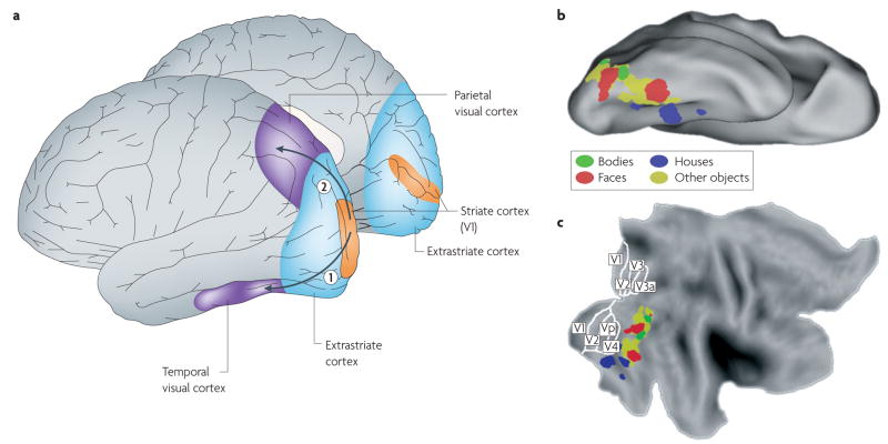
a | The location of visual regions in the human cortex, including the primary visual cortex (area V1 in the striate cortex) and the extrastriate cortex in the occipital lobe, and the traditional distinction into two visual cortical pathways that start in area V1 and extend into the temporal lobe (the ventral ‘what’ or ‘object-vision’ pathway (1)) or into the parietal lobe (the dorsal ‘where’ pathway (2)). b,c | Ventral pathway regions in one individual that were activated significantly at the voxel level (P < 0.0001, uncorrected) in the following contrasts: bodies > faces + houses (shown in green); faces > bodies + houses (shown in red); houses > bodies + faces (shown in blue). In addition, the yellow areas represent the regions that, in a group of people (n = 9), activated significantly in the contrast: intact objects > scrambled objects. All data were processed using SPM5 (Wellcome Department of Cognitive Neurology, London). Data are shown on top of the PALS human atlas (using CARET software138,139) in a ventral view of the inflated cortical surface (b) and in a flattened view of the cortical surface (c). The partitioning of retinotopic areas in the striate and extrastriate cortex is shown as included in the PALS atlas.
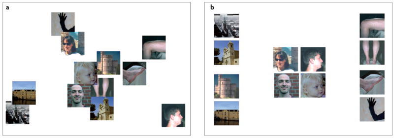
a | A two-dimensional representation of the physical differences that exist between twelve images, as quantified by the luminance difference between corresponding pixels, summed across pixels. b | A two-dimensional representation of the perceived differences between the same twelve images, as indicated by a human observer. Note that there is no correspondence between this higher-order mental space and the physical space of part a. The two-dimensional representations were obtained by applying non-metric multidimensional scaling to the matrices, with pair-wise physical (a) and perceived (b) differences.
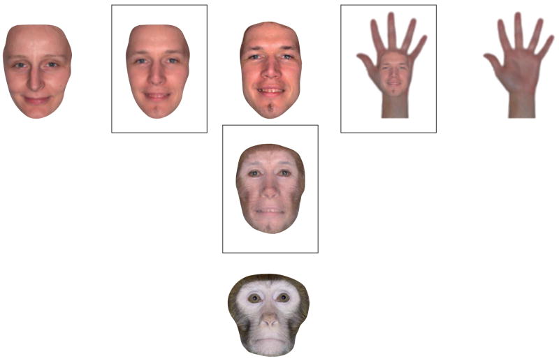
This figure shows morphed images (in squares) that are, respectively, combinations of two human faces (left); a human face and a monkey face (bottom); and a human face and a human hand (right). For two human faces, or even for the faces of members of different species, corresponding points in the two figures can be easily found. However, it is not straightforward to identify corresponding points on objects that are more distant in object space, such as a face and a hand.
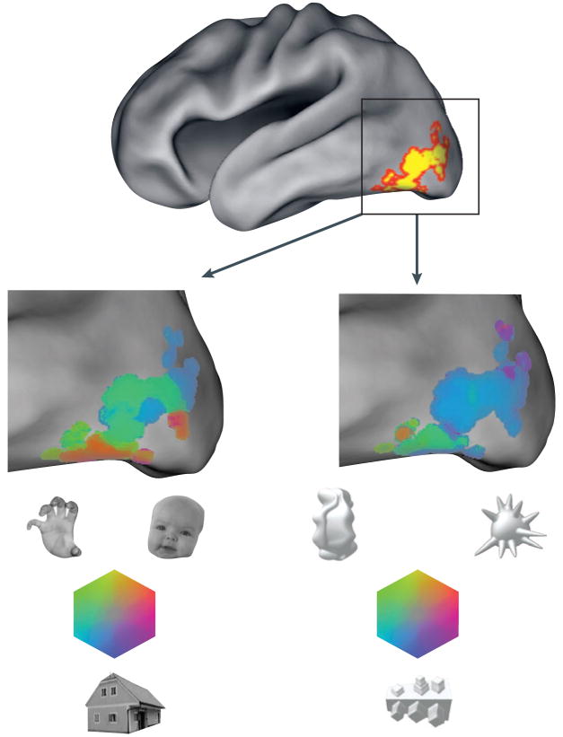
Functional specificity in the lateral occipital and ventral occipitotemporal cortex (the ‘lateral occipital complex’, as defined by the contrast: intact objects > scrambled objects) is shown by a colour map for two sets of stimuli: three familiar objects (faces, body parts and houses; left-hand enlargement) and three novel objects (right-hand enlargement). Colour saturation represents the amount of selectivity; hue represents which object class is preferred. It is important to note that the colour scale is not given a threshold for statistical significance, and few individual voxels show significant specificity for the novel objects. Nevertheless, the pattern of selectivity across many voxels is replicable. In this individual the spatial correlation across voxels between independent subsets of the data (‘odd’ and ‘even’ runs) was 0.78 for familiar objects and 0.52 for novel objects.
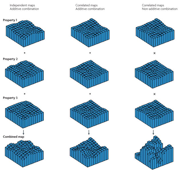
Each plot in the top three rows shows the response profile of a hypothetical voxel set to a particular functional property. The bottom row illustrates different possibilities for the combination of these overlapping maps. If the individual maps are uncorrelated and their integration is additive (left-hand column), the resulting combined selectivity profile will be similar to those of the individual properties. If the individual maps are correlated and additively combined (middle column), the joint presence of all three features will lead to a more selective reponse profile. If the individual maps are correlated and combined nonlinearly (here, by multiplication; right-hand column), the resulting selectivity profile will be pronounced, with subsets of the voxel space responding strongly to the joint presence of two or more individual properties.
References
-
- Grill-Spector K, Kourtzi Z, Kanwisher N. The lateral occipital complex and its role in object recognition. Vision Res. 2001;41:1409–1422. - PubMed
-
- Kanwisher N, Woods RP, Iacoboni M, Mazziotta JC. A locus in human extrastriate cortex for visual shape analysis. J Cogn Neurosci. 1997;9:133–142. - PubMed
Publication types
MeSH terms
Grants and funding
LinkOut - more resources
Full Text Sources
Medical
Miscellaneous
