

Denatured collagen modulates the phenotype of normal and wounded human skin equivalents
- PMID: 18200055
- PMCID: PMC2810554
- DOI: 10.1038/sj.jid.5701240
Denatured collagen modulates the phenotype of normal and wounded human skin equivalents
Abstract
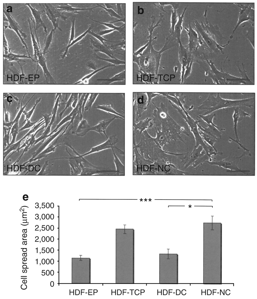
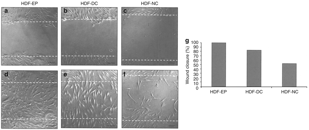
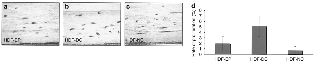
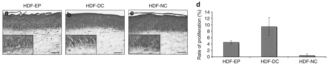
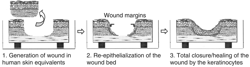
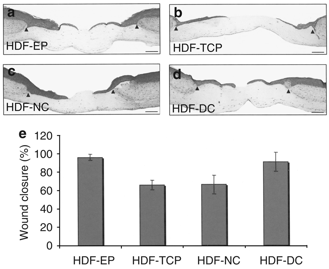
Epithelial-mesenchymal interactions are known to play an important role in modulating homeostasis and repair. However, it remains unclear how the composition of the extracellular matrix may regulate the ability of dermal fibroblasts to engage in such cross talk. To address this, we studied how fibroblast phenotype was linked to the behavior of normal and wounded human skin equivalents (HSE) by comparing human dermal fibroblasts (HDF) incorporated into the three-dimensional tissues to those extensively cultivated in two-dimensional (2D) monolayer culture on denatured collagen (DC) matrix, native collagen, or tissue culture plastic before incorporation into HSEs. We first established that prolonged passage and growth of HDF on DC increased their migratory potential in a 2D monolayer culture. When HDF variants were grown in HSEs, we found that extended passage on DC and incorporation of DC directly into the collagen gel enhanced proliferation of both HDF and basal keratinocytes in HSEs. By adapting HSEs to study wound reepithelialization, we found that the extended passage of HDF on DC accelerated the rate of wound healing by 38%. Thus, extensive ex vivo expansion on DC was able to modify the phenotype of skin fibroblasts by augmenting their reparative properties in skin-like HSEs.
Conflict of interest statement
The authors state no conflict of interest.
Figures

Similar articles
-
Three-dimensional human tissue models of wounded skin.Methods Mol Biol. 2010;585:345-59. doi: 10.1007/978-1-60761-380-0_24. Methods Mol Biol. 2010. PMID: 19908015 Free PMC article.
-
Effect of hyperbaric oxygen on human skin cells in culture and in human dermal and skin equivalents.Wound Repair Regen. 1999 Jan-Feb;7(1):53-64. doi: 10.1046/j.1524-475x.1999.00053.x. Wound Repair Regen. 1999. PMID: 10231506
-
Development of a Full-Thickness Human Skin Equivalent In Vitro Model Derived from TERT-Immortalized Keratinocytes and Fibroblasts.Tissue Eng Part A. 2015 Sep;21(17-18):2448-59. doi: 10.1089/ten.TEA.2015.0139. Epub 2015 Aug 3. Tissue Eng Part A. 2015. PMID: 26135533 Free PMC article.
-
Understanding experimental biology of skin equivalent: from laboratory to clinical use in patients with burns and chronic wounds.Am J Surg. 2004 May;187(5A):29S-33S. doi: 10.1016/S0002-9610(03)00301-5. Am J Surg. 2004. PMID: 15147989 Review.
-
Keratinocyte-fibroblast interactions in wound healing.J Invest Dermatol. 2007 May;127(5):998-1008. doi: 10.1038/sj.jid.5700786. J Invest Dermatol. 2007. PMID: 17435785 Review.
Cited by
-
Three-dimensional human tissue models of wounded skin.Methods Mol Biol. 2010;585:345-59. doi: 10.1007/978-1-60761-380-0_24. Methods Mol Biol. 2010. PMID: 19908015 Free PMC article.
-
Activated protein C inhibits proliferation and tumor necrosis factor α-stimulated activation of p38, c-Jun NH2-terminal kinase (JNK) and Akt in rheumatoid synovial fibroblasts.Mol Med. 2013 Oct 24;19(1):324-31. doi: 10.2119/molmed.2013.00034. Mol Med. 2013. PMID: 24096826 Free PMC article.
-
Fibroblasts derived from human embryonic stem cells direct development and repair of 3D human skin equivalents.Stem Cell Res Ther. 2011 Feb 21;2(1):10. doi: 10.1186/scrt51. Stem Cell Res Ther. 2011. PMID: 21338517 Free PMC article.
-
Primary cultured fibroblasts derived from patients with chronic wounds: a methodology to produce human cell lines and test putative growth factor therapy such as GMCSF.J Transl Med. 2008 Dec 1;6:75. doi: 10.1186/1479-5876-6-75. J Transl Med. 2008. PMID: 19046453 Free PMC article.
-
Aegyptin displays high-affinity for the von Willebrand factor binding site (RGQOGVMGF) in collagen and inhibits carotid thrombus formation in vivo.FEBS J. 2010 Jan;277(2):413-27. doi: 10.1111/j.1742-4658.2009.07494.x. Epub 2009 Dec 15. FEBS J. 2010. PMID: 20015075 Free PMC article.
References
-
- Abraham LC, Vorrasi J, Kaplan DL. Impact of collagen structure on matrix trafficking by human fibroblasts. J Biomed Mater Res A. 2004;70:39–48. - PubMed
-
- Ali-Bahar M, Bauer B, Tredget EE, Ghahary A. Dermal fibroblasts from different layers of human skin are heterogeneous in expression of collagenase and types I and III procollagen mRNA. Wound Repair Regen. 2004;12:175–182. - PubMed
-
- Andriani F, Margulis A, Lin N, Griffey S, Garlick JA. Analysis of microenvironmental factors contributing to basement membrane assembly and normalized epidermal phenotype. J Invest Dermatol. 2003;120:923–931. - PubMed
-
- Aumailley M, Mann K, von der MH, Timpl R. Cell attachment properties of collagen type VI and Arg-Gly-Asp dependent binding to its alpha 2(VI) and alpha 3(VI) chains. Exp Cell Res. 1989;181:463–474. - PubMed
Publication types
MeSH terms
Substances
Grants and funding
LinkOut - more resources
Full Text Sources
Other Literature Sources
