Review
. 2008 Sep;97(9):3518-90.
doi: 10.1002/jps.21270.

Nanovehicular intracellular delivery systems

Affiliations
Review

Nanovehicular intracellular delivery systems

Ales Prokop et al. J Pharm Sci. 2008 Sep.

Abstract

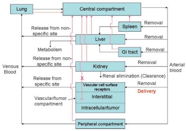
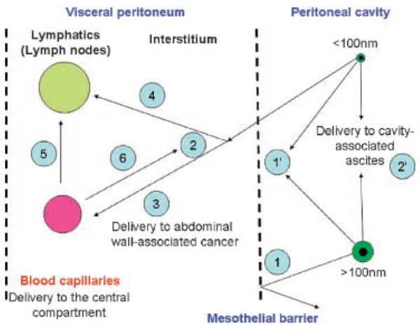
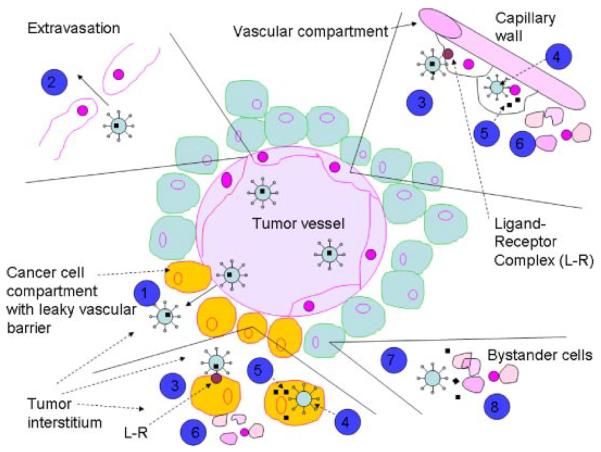
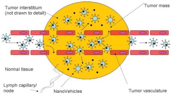
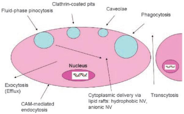
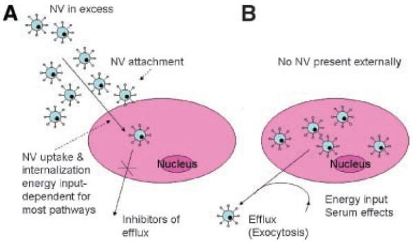
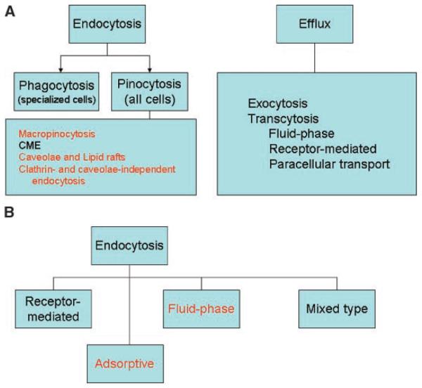
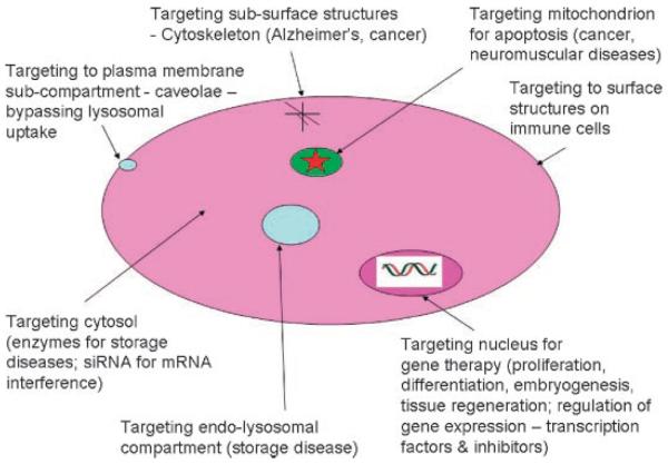
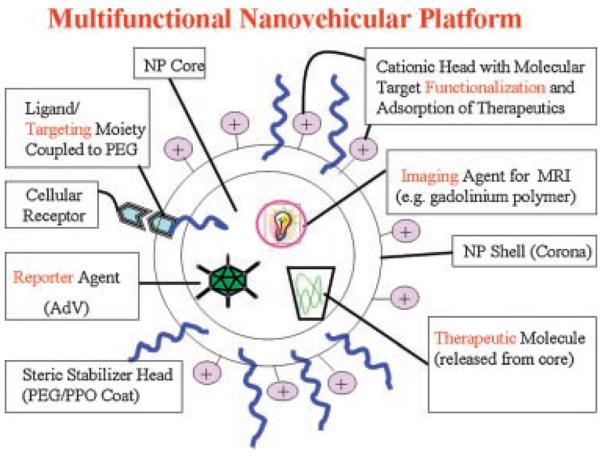
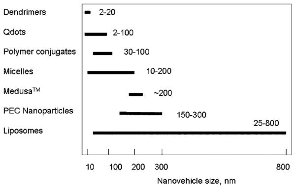
This article provides an overview of principles and barriers relevant to intracellular drug and gene transport, accumulation and retention (collectively called as drug delivery) by means of nanovehicles (NV). The aim is to deliver a cargo to a particular intracellular site, if possible, to exert a local action. Some of the principles discussed in this article apply to noncolloidal drugs that are not permeable to the plasma membrane or to the blood-brain barrier. NV are defined as a wide range of nanosized particles leading to colloidal objects which are capable of entering cells and tissues and delivering a cargo intracelullarly. Different localization and targeting means are discussed. Limited discussion on pharmacokinetics and pharmacodynamics is also presented. NVs are contrasted to micro-delivery and current nanotechnologies which are already in commercial use. Newer developments in NV technologies are outlined and future applications are stressed. We also briefly review the existing modeling tools and approaches to quantitatively describe the behavior of targeted NV within the vascular and tumor compartments, an area of particular importance. While we list "elementary" phenomena related to different level of complexity of delivery to cancer, we also stress importance of multi-scale modeling and bottom-up systems biology approach.


Figures

Figure 1
Figure 1
Systemic administration of a drug-nanovehicle into the central (blood-lymph) compartment. In pharmacokinetic models each organ could be composed if multiple compartments, which reflect the anatomy/morphology of the organ. Red arrow symbolizes the lymph drainage from each organ(s). Note that the tumor lymph drainage is often impaired. For peripheral compartment, most organs are drained.
Figure 2
Figure 2
Major elements of the intraperitoneal drug delivery. Nanovehicles > 100 nm are rejected (1), but smaller NP can pass from the peritoneal cavity and through the mesothelium into the peritoneal interstitium (2). (1′) represents a delivery to mesotheliomas. (2′) denotes interstitial uptake (by abdominal tumors and ascites). The peritoneal membrane is an idealized partition barrier with heterogeneous sieving characteristics. By diffusion and convection, NP can enter discrete blood (3) and lymph capillaries (4) within the interstitium. Macromolecules and nanovehicles could also diffuse from the blood compartment to the lymphatic (5) or interstitial (6) compartments. Interstitium (peritoneal tissue) is dense network composed of collagenous, glycoprotein, and proteoglycan material. Adapted from Flessner.
Figure 3
Figure 3
Schematic diagram of mechanism of targeted nanovehicle delivery of a therapeutic drug to vascular compartment and into solid tumors. Vascular targeting agents often exhibit an affinity to both endothelial and tumor cells. Targeted nanovehicles, endowed with a specific targeting ligand on their periphery, accumulate passively in tumor tissue because EPR (enhanced permeability and retention) effect and preferential extravasation (1) from tumor vessels. Endothelial cells are shed (partially) from the lining of tumor blood vessels, exposing underlying tumor cells. Consequently, increased vascular permeability of vascular tissue (leaky endothelium) enables nanovehicles to extravasate and reach the tumor interstitial fluid. This passive and nonspecific process of nanovehicle extravasation is statistically improved by the prolonged residence time of nanovehicles in circulation and repeated passages through the tumor microvascular bed. Nanovehicles with engineered (PEG and other technologies) long-circulating properties increase the number of passages through the tumor microvasculature. However, except for rare instances, tumor cells are not directly exposed to the blood stream. Therefore, for an intravascular targeting device to access the tumor cell, it must first cross the vascular endothelium and diffuse into the interstitial fluid, via extravasation (2). Extravasated nanovehicles then attach to cancer cells (3) and are taken up (internalized) by tumor tissue (4). Likewise, an attachment and internalization of nanovehicles may happen with endothelial cells because of specific vascular targeting agent. Subsequently to internalization, intracellular drug cytosolic release (5) occurs, followed by direct killing of tumor and endothelial cells (6). Once nanovehicles have penetrated the tumor interstitial fluid, binding of targeted ligand-endowed nanovehicles may occur vigorously, shifting the intratumor distribution from the extracellular compartment to the tumor cell intracellular compartment. This shift could be several times higher for targeted nanovehicles as compared to nontargeted ones. Also, the recirculation of nanovehicles within the blood compartment will be considerably reduced for nanovehicles with specific-binding affinity to tumor cell receptors. Because of limited diffusion capacity of nanovehicles within the interstitial space, binding is likely to be limited to the tumor cells in closest vicinity to blood vessels. In addition, the nanovehicles which fail to bind to tumor cells will reside in the extracellular (interstitial) space. Upon their destabilization, they slowly release (7) their drug content into the interstitial space which will eventually diffuse to nearby cancer cells and bystander cells (8) exerting a cytotoxic effect. Obviously, there will always be a combination of in situ release from an extracellular nanovehicle depot and intracellular release from internalized nanovehicles. Therefore, the theoretical advantages of targeted nanovehicles over the nontargeted are related to a shift of nanovehicle distribution to the tumor cell compartment, delivery of nanovehicular contents to an intracellular tumor compartment in nanovehicle-associated form, and, possibly, prolonged nanovehicle retention in the tumor (provided with a proper PEG chemistry). Adapted from Park et al. and Gabizon et al.
Figure 4
Figure 4
Schematic of EPR (enhanced permeability and retention) effect in solid tumors: nanovehicles passively target to vasculature and extravasate through fenestrated tumor vasculature. Nanovehicles accumulate in tumor tissue because of their extended circulation time in conjunction with preferential extravasation from tumor vessels (EPR effect) and lack of lymphatic drainage. This passive targeting process facilitates tumor tissue binding, followed by uptake (internalization). Resulting is intracellular drug release for drug action and cell killing. Nanovehicles which fail to bind to tumor cells will reside in the extracellular (interstitial) space, where they eventually become destabilized because of enzymatic and phagocytic attack. This results in extracellular drug release for eventual diffusion to nearby tumor cells and bystander cell. Normal blood vessels have a tight endothelium. Adapted from van Vlerken and Amiji.
Figure 5
Figure 5
Schematic representation of uptake and exocytosis routes of nanovehicles. Redrawn from Panyam and Labhasetwar. Not drawn to scale.
Figure 6
Figure 6
Kinetic considerations at nanovehicle uptake and exocytosis in vitro. The overall fate could be traced by flow cytometry (FACS) following the cell detachment via trypsin or EDTA (ethylenediamine tetraacetic acid). Nanovehicles must be present in medium in excess to be internalized. The efflux could be inhibited by specific inhibitors. The quantity of nanovehicles could be distinguished from total uptake by fluorescence quenching by Trypan Blue (A). When resuspended in fresh media without nanovehicles, they can efflux to the environment. This process requires energy and presence of serum (B).
Figure 7
Figure 7
Nanovehicle uptake and efflux routes. (A) Mechanistic view (pathways in red lettering indicate the preferred routes of uptake, bypassing endosomal trafficking, but not very efficient in terms of uptake rate). (B) Kinetic view (preferred routes). Adapted from Khalil et al.
Figure 8
Figure 8
Some possible disease intracellular targets.
Figure 9
Figure 9
Multifunctional nanovehicle platform. NV (nanovehicle) core contains a therapeutic molecule, which can be released from NP (or delivered intracellularly, following NV internalization); it could also possess a molecular imaging agent—for example, a magnetic resonance imaging (MRI) agent, and reporter of efficacy—luminescence/imaging reporter agent (adV). The corona is typically cationically charged, with primary amino groups which serve as a electrostatic stabilizer, and could be used to functionalize the NV surface, for example, with a targeting moiety (e.g., a peptide with an affinity to HSPG molecules); a therapeutic (peptide, protein) could be adsorbed onto the NP surface via the electrostatic interactions to provide a suitable solid-phase signal; a targeting ligand could also be coupled to a terminal end-group of PEG molecule (Pluronic F-68), serving, in the first place, as a steric stabilizer.
Figure 10
Figure 10
Typical size range of representative examples of nanovehicles. Note a lower limit on liposomes (vehicle stability), nanoparticles (condensation limit) and upper limit on dendrimers, Qdots, and polymer conjugates (due to their chemistry/structure). Larger sizes may not be optimal for accessing/delivery of certain cells and tissues.

References

    1. Kostarelos K. Rational design and engineering of delivery systems for therapeutics: Biomedical exercises in colloid and surface science. Adv Colloid Interface Sci. 2003;106:147–168. - PubMed
    1. Moses MA, Brem H, Langer R. Advancing the field of drug delivery: Taking aim at cancer. Cancer Cell. 2003;4:337–341. - PubMed
    1. Shargel L, Yu AB. Applied biopharmaceutics & pharmacokinetics. 4th edition McGraw-Hill; New York: 1999.
    1. van de Waterbeemd H, Gifford E. ADMET in silico modelling: Towards prediction paradise? Nat Rev Drug Discov. 2003;2:192–204. - PubMed
    1. Okada H, Toguchi H. Biodegradable microspheres in drug delivery. Crit Rev Ther Drug Carrier Syst. 1995;12:1–99. - PubMed

Publication types