. 2008 May;53(5):405-15.
doi: 10.1016/j.archoralbio.2007.11.014. Epub 2008 Jan 16.

Cytomegalovirus inhibition of embryonic mouse tooth development: a model of the human amelogenesis imperfecta phenocopy

Affiliations

Cytomegalovirus inhibition of embryonic mouse tooth development: a model of the human amelogenesis imperfecta phenocopy

Tina Jaskoll et al. Arch Oral Biol. 2008 May.

Abstract

Objective: Cytomegalovirus (CMV) is one of the most common causes of major birth defects in humans. Of the approximately 8400 children born each year in the U.S. with CMV-induced birth defects, more than 1/3 of these children exhibit hypoplasia and hypocalcification of tooth enamel. Our objective was to initiate the investigation of the pathogenesis of CMV-induced tooth defects.

Design: Mouse Cap stage mandibular first molars were infected with mouse CMV (mCMV) in vitro in a chemically-defined organ culture system and analysed utilising histological and immunolocalisation methodologies. The antiviral, acyclovir, was used to inhibit mCMV replication and comparisons made between mCMV-infected and acyclovir-treated, mCMV-infected teeth.

Results: Active infection of Cap stage molars for up to 15 days in vitro results in smaller, developmentally-delayed and dysmorphic molars characterised by shallow, broad and misshapen cusps, infected and affected dental papilla mesenchyme, poorly differentiated odontoblasts and ameloblasts, and no dentin matrix. Initial protein localisation studies suggest that the pathogenesis is mediated through NF-kappaB signaling and that there appears to be an unusual interaction between abnormal mesenchymal cells and surrounding matrix. Rescue with acyclovir indicates that mCMV replication is necessary to initiate and sustain progressive tooth dysmorphogenesis.

Conclusions: Our results indicate that mCMV-induced changes in signaling pathways severely delays, but does not completely interrupt, tooth morphogenesis. Importantly, our results demonstrate that this well-defined embryonic mouse organ culture system can be utilised to delineate the molecular mechanism underlying the CMV-induced tooth defects that characterise the amelogenesis imperfecta phenocopy seen in many CMV-infected children.


Figures
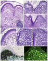
Fig. 1
Fig. 1
mCMV-induced histopathology and viral distribution. (A–F) Histological analysis of control (A and D) and mCMV-infected (B and C, E and F) E15 Cap stage first mandibular molars cultured in serumless, chemically-defined medium. (A and D) After 12 days in culture, control molars have undergone cuspal morphogenesis and reached the Bell stage, with polarised odontoblast (o) and ameloblasts (a), as well as dentin matrix (d), being seen. dp, dental papilla. (Insert A) Starting E15 Cap stage mandibular first molar. (B and E) After 12 days in culture, the mCMV-infected molars are markedly smaller, developmentally-delayed, and severely abnormal, having only achieving the Early Bell stage. mCMV infection results in shallow, broad and misshapen cusps composed of abnormally short, undifferentiated dental epithelium (arrow), disorganised undifferentiated odontoblasts (double arrowheads), and no dentin matrix. The dental papilla is composed of clusters of large basophilic, pleiomorphic infected [with viral inclusion bodies (insert)] and affected cells being seen (double arrows). (C and F) After 15 days of infection, the smaller, underdeveloped and abnormal tooth phenotype persists. The presence of aligned (but not polarised) preodontoblasts (arrowhead), secreted predentin matrix (white arrow), and elongated (but not polarised) epithelia (white arrowhead) indicates developmental advancement. (Insert F) Viral inclusion bodies. (G and H) mCMV distribution in E15 + 12 tooth organs. Expression of β-galactosidase staining of lacZ (mCMV) (G) and viral IE1 protein (H) demonstrates the presence of viral infection throughout dental papilla EM cells but not in dental epithelia (e). Bar (A–C), 30 μm; (A insert), 60 μm; (D–F, H), 20 μm; (E and F insert), 12 μm; (G), 50 μm.
Fig. 2
Fig. 2
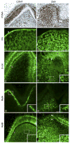
Characterisation of mCMV-induced cellular changes. (A and B) Cell proliferation. Cell proliferation was determined by the distribution of PCNA-positive nuclei (brown colour). In control molars (A), PCNA-positive nuclei are seen in ameloblasts (a) and, to a lesser extent, in dental papilla (dp) mesenchyme. Note the absence of PCNA-positive nuclei in polarised odontoblasts (o) in the cuspal tip, indicating differentiation. With mCMV infection (B), PCNA-positive nuclei are seen throughout the cytomegalic mesenchymal cell population (*) and, to a lesser extent, in short, nonpolarised epithelial cells (double arrows). (C and D) FN distribution. In control molars (C), FN is strongly immunolocalised in basement membrane (white arrowhead) and weakly throughout the dental papilla extracellular matrix. With mCMV infection (D), FN intensely surrounds individual cytomegalic dental papilla cells (white asterisks) and is relatively absent from the basement membrane. (E and F) β-Catenin distribution. In control molars (E), β-catenin is seen in the apical and basal regions (arrows) of polarised ameloblasts and more weakly in aligned odontoblasts. In mCMV-infected molars (F), β-catenin is strongly accumulated in the cytoplasm of dental papilla mesenchymal (insert) and short, nonpolarised epithelial (e) cells (white arrows); β-catenin is also detected in cell membranes. (G and H) RelA distribution. In control molars (G), polarised odontoblasts exhibit nuclear-localised RelA (insert). With mCMV infection (H), RelA is seen in the cytoplasm of centrally-localised cytomegalic dental papilla cells (insert) and is absent from presumptive odontoblasts. (I and J) RelB distribution. In control molars (I), RelB is seen in the apical and basal regions of polarised ameloblasts (black arrowheads). A similar cytoplasmic distribution pattern (black arrowheads) is seen in the mCMV-infected epithelia (D) which consists of nonpolarised cuboidal epithelial cells (white arrowheads). Note that mCMV infection induced a marked increase in RelB immunostain in the cytoplasm of centrally-localised dental papilla mesenchyme (insert D) compared to control (insert C). Bar (A and B, E–J), 20 μm; (C and D), 10 μm. (F–J inserts), 13 μm.
Fig. 3
Fig. 3
Acyclovir treatment ameliorates mCMV-induced pathology. (A–D) β-Galactosidase (lacZ) expression. LacZ is detected in EM dental papilla cells in mCMV-infected tooth organs (A and B) and was absent from acyclovir-treated, mCMV-infected (CMV + Acy) tooth organs (C and D). (E–G) Histological sections of control (E), mCMV-infected (F) and mCMV + Acy (G) molars. mCMV infection induces abnormal tooth development (F), characterised by unaligned, disorganised odontoblasts (arrow), undifferentiated ameloblasts, and abnormal mesenchymal cellularity. In contrast, acyclovir-treated, mCMV-infected molar phenotypes (G) are similar to that seen in control (E). Note, the presence of aligned, polarised odontoblasts (double arrows) and ameloblasts (double arrowheads), increased dentin matrix deposition (arrowhead), and dental papilla cells exhibiting a normal, fibroblastic appearance. Bar (A and C), 30 μm; (B, D–G), 20 μm.

Similar articles

Cited by

References

    1. Pass RF. Cytomegalovirus. In: Knipe DM, Howley PM, Griffin DE, Lamb RA, Martin MA, Roizman B, et al., editors. Fields virology. 4th. Philadelphia: Lippincott Williams & Wilkins; 2001. pp. 2675–705.
    1. Dollard SC, Grosse SD, Ross DS. New estimates of the prevalence of neurological and sensory sequelae and mortality associated with congenital cytomegalovirus infection. Rev Med Virol. 2007;17:355–63. - PubMed
    1. Ross DS, Dollard SC, Victor M, Sumartojo E, Cannon MJ. The epidemiology and prevention of congenital cytomegalovirus infection and disease: activities of the Centers for Disease Control and Prevention Workgroup. J Womens Health (Larchmt) 2006;15:224–9. - PubMed
    1. Stagno S, Pass RF, Dworsky ME, Alford CA. Congenital and perinatal cytomegalovirus infections. Semin Perinatol. 1983;7:31–42. - PubMed
    1. Stagno S, Pass RF, Dworsky ME, Britt WJ, Alford CA. Congenital and perinatal cytomegalovirus infections: clinical characteristics and pathogenic factors. Birth Defects Orig Article Ser. 1984;20:65–85. - PubMed

Publication types

MeSH terms