. 2008 Mar 10;314(5):1094-104.
doi: 10.1016/j.yexcr.2007.12.010. Epub 2007 Dec 23.

Connective tissue growth factor/CCN2-null mouse embryonic fibroblasts retain intact transforming growth factor-beta responsiveness

Affiliations

Connective tissue growth factor/CCN2-null mouse embryonic fibroblasts retain intact transforming growth factor-beta responsiveness

Yasuji Mori et al. Exp Cell Res. .

Abstract

Background: The matricellular protein connective tissue growth factor (CCN2) has been implicated in pathological fibrosis, but its physiologic role remains elusive. In vitro, transforming growth factor-beta (TGF-beta) induces CCN2 expression in mesenchymal cells. Because CCN2 can enhance profibrotic responses elicited by TGF-beta, it has been proposed that CCN2 functions as an essential downstream signaling mediator for TGF-beta. To explore this notion, we characterized TGF-beta-induced activation of fibroblasts from CCN2-null (CCN2(-/-)) mouse embryos.

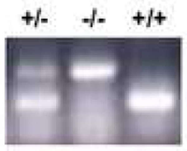
Methods: The regulation of CCN2 expression was examined in vivo in a model of fibrosis induced by bleomycin. Cellular TGF-beta signal transduction and regulation of collagen gene expression were examined in CCN2(-/-) MEFs by immunohistochemistry, Northern, Western and RT-PCR analysis, immunocytochemistry and transient transfection assays.

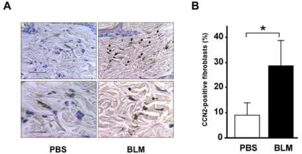
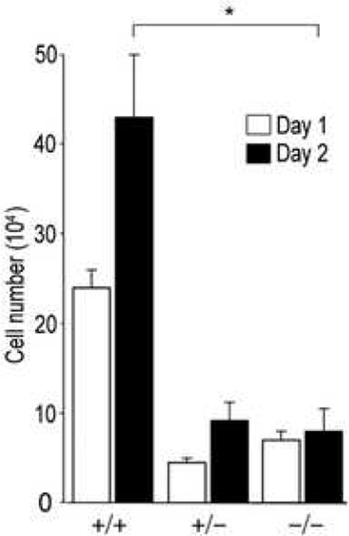
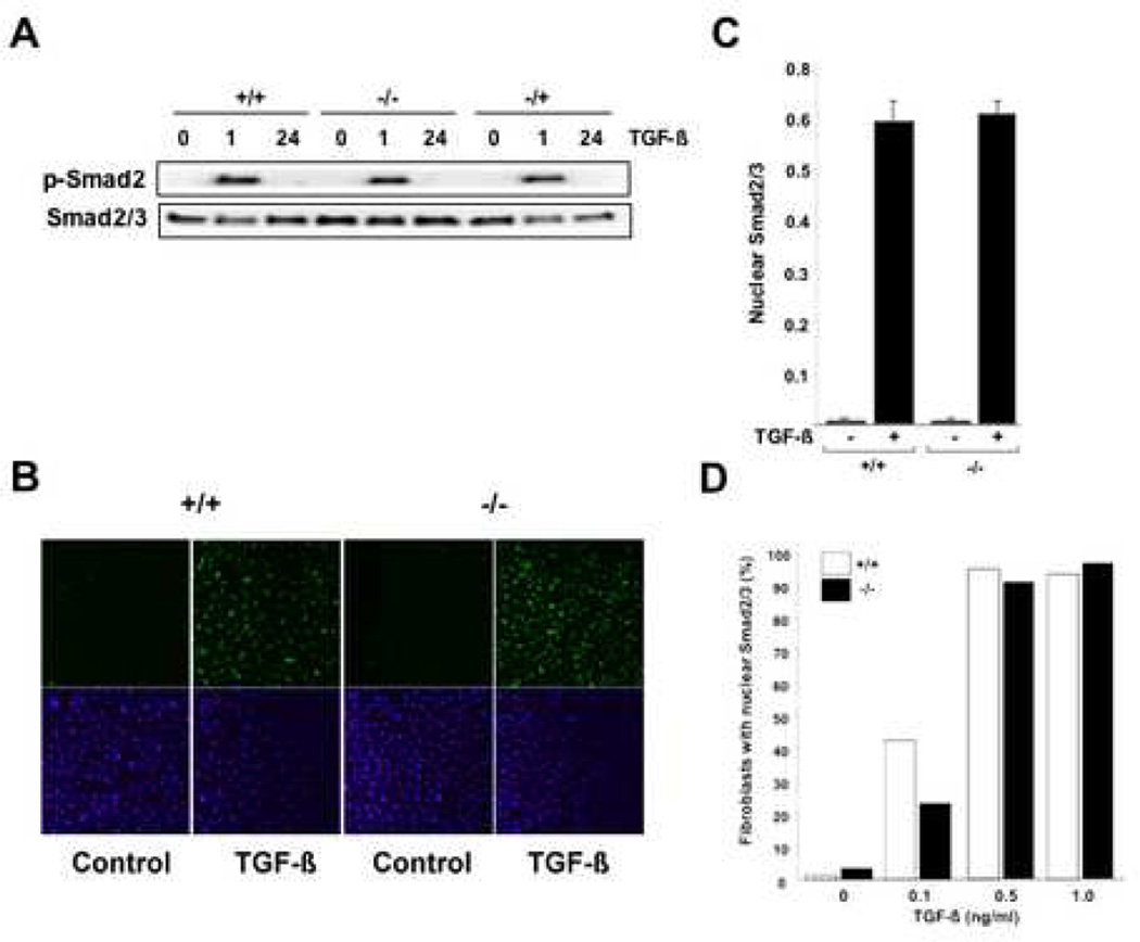
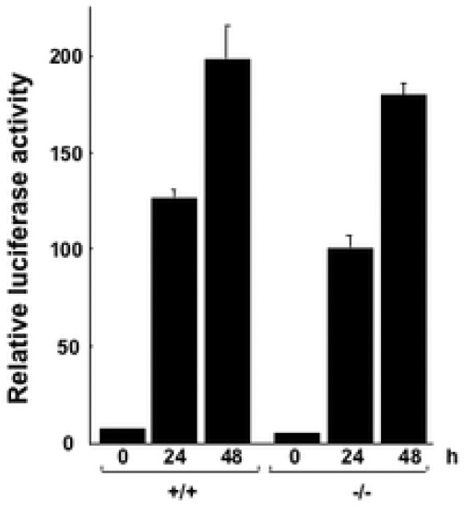
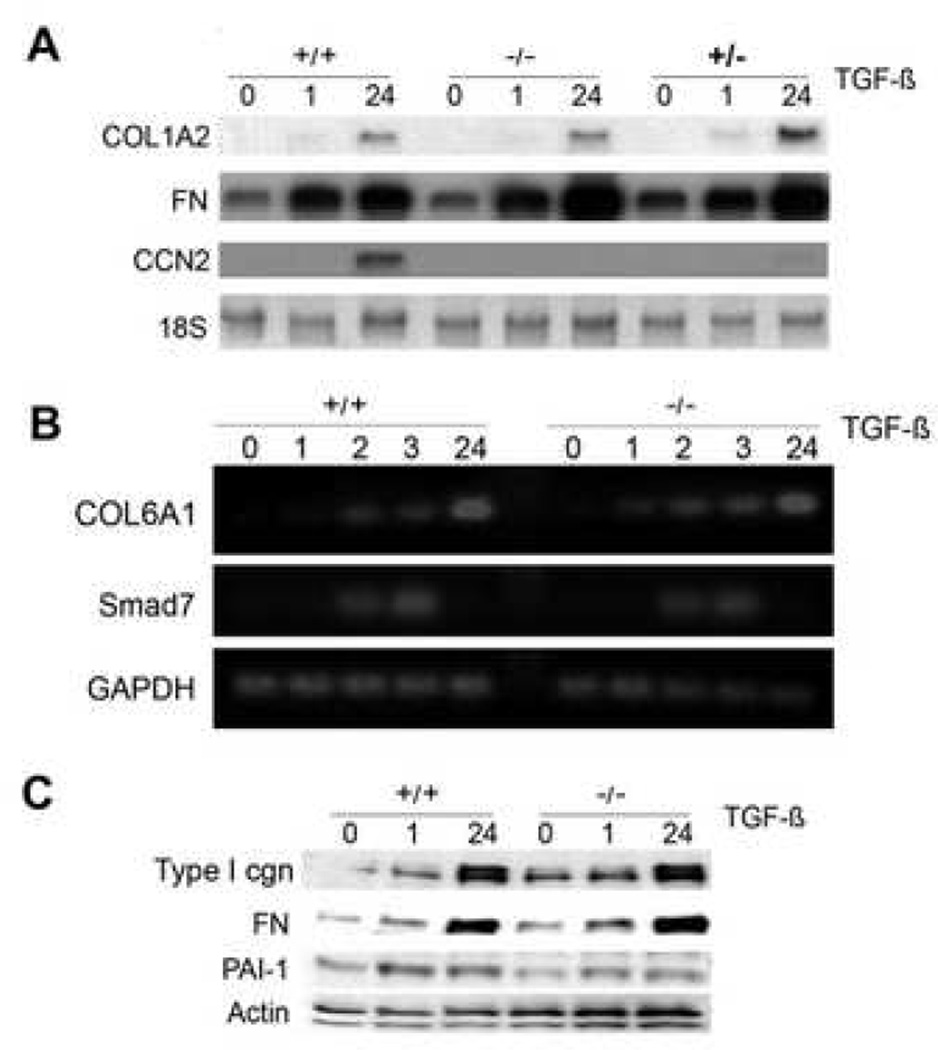
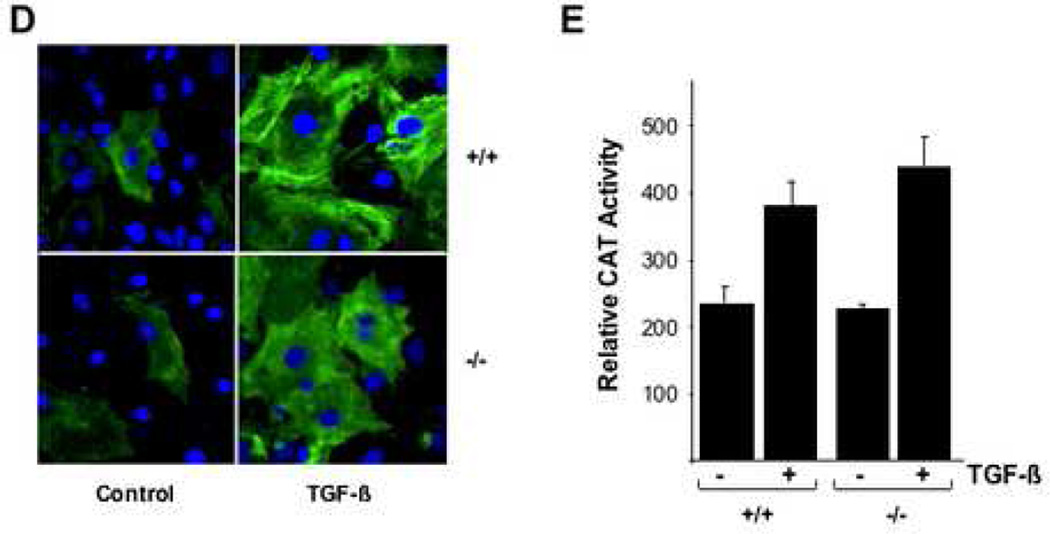
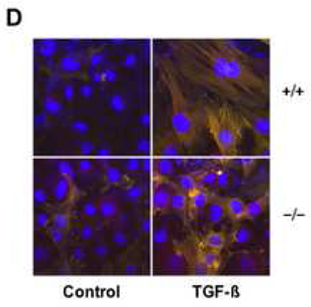
Results: Bleomycin-induced skin fibrosis in the mouse was associated with substantial CCN2 up-regulation in lesional fibroblasts. Whereas in vitro proliferation rate of CCN2(-/-) MEFs was markedly reduced compared to wild type MEFs, TGF-beta-induced activation of the Smad pathways, including Smad2 phosphorylation, Smad2/3 and Smad4 nuclear accumulation and Smad-dependent transcriptional responses, were unaffected by loss of CCN2. The stimulation of COL1A2 and fibronectin mRNA expression and promoter activity, and of corresponding protein levels, showed comparable time and dose-response in wild type and CCN2(-/-) MEFs, whereas stimulation of alpha smooth muscle actin and myofibroblast transdifferentiation showed subtle impairment in MEFs lacking CCN2.

Conclusion: Whereas endogenous CCN2 plays a role in regulation of proliferation and TGF-beta-induced myofibroblast transdifferentiation, it appears to be dispensable for Smad-dependent stimulation of collagen and extracellular matrix synthesis in murine embryonic fibroblasts.


Figures

Figure 1
Figure 1. Up-regulation of CCN2 expression in lesional dermis
C57BL/6 mice (6–8 week old) received daily s.c. injections of bleomycin or PBS in parallel. At 21 days, lesional skin was harvested and tissue sections were examined by immunohistochemistry using antibodies to CCN2. A. Representative photomicrographs. Arrows indicate positively stained fibroblasts. Bars, 50 µm (upper panels), 20 µm (lower panels). B. The proportion of CCN2-positive fibroblastic cells. The results are expressed as means ± SD of percentage of CCN2-positive fibroblasts from six independent microscopic fields; p<0.05.
Figure 2
Figure 2. PCR analysis of CCN2-null MEFs
Genomic DNA was extracted from CCN2−/−, CCN2−/+ and CCN2−/− MEFs and used for PCR analysis with primers specific for CCN2.
Figure 3
Figure 3. Murine embryonic fibroblast proliferation
Fibroblasts from CCN2−/−, CCN2−/+ and CCN2+/+ embryos were seeded in 6-well plates, and after 1 or 2 d incubation, cell numbers were determined. Results from a representative experiment (means ± SEM from triplicate determinations); p<0.05. Viability was >95%.
Figure 4
Figure 4. Smad activation in CCN2−/− MEFs
Confluent cultures of CCN2−/−, CCN2−/+ and CCN2−/− MEFs in parallel were incubated with TGF-β for indicated periods. A. Whole cell lysates were prepared and subjected to Western analysis. Representative autoradiograms are shown. B. Cultures were incubated with TGF-β for 60 min, fixed and examined by immunocytochemistry and confocal microscopy using antibodies to Smad2/3. Nuclei were identified by DAPI (blue). Representative images are shown. C. The proportion of MEFs showing predominantly nuclear Smad2/3 was determined as described in Materials and Methods. Results are expressed as means ± SEM from 5 independent determinations from three different microscopic fields. D. Comparable dose-response of TGF-β-induced Smad2/3 nuclear accumulation in CCN2−/− and CCN2−/− MEFs. Fibroblasts were categorized as showing predominantly nuclear Smad2/3 by a blinded observer.
Figure 5
Figure 5. SMAD-dependent transcriptional activity in CCN2−/− MEFs
Subconfluent cultures of wild type and CCN2−/− MEFs were transiently transfected with p[SBE]4-luc and following 48 h incubation with TGF-β luciferase activities were determined. The results, normalized by renilla luciferase activity to correct for small variations in transfection efficiency between samples, represent the means ± SEM from triplicate determinations. Representative results from three experiments.
Figure 6
Figure 6. Regulation of ECM gene expression by TGF-β in CCN2−/− MEFs
Subconfluent cultures of wild type, CCN2+/− and CCN2−/− MEFs were incubated with TGF-β for the indicated periods, and mRNA levels were examined by A. Northern analysis; or B. RT-PCR analysis. Representative results shown. C. Whole cell lysates were subjected to Western analysis. D. Confocal microscopy was performed in confluent cultures incubated with TGF-β for 48 h and stained with antibodies to Type I collagen (green color). E. Cultures were transiently transfected with 772COL1A2-CAT, and following 48 h incubation with TGF-β, cell lysates were assayed for their CAT activities. The results, normalized by renilla luciferase activity to correct for small variations in transfection efficiency between samples, represent the means ± SEM from triplicate determinations.
Figure 7
Figure 7. Induction of myofibroblasts differentiation in CCN2−/− MEFs
A. Subconfluent cultures of wild type and CCN2−/− MEFs were incubated in parallel with TGF-β for 48 h, and whole cell lysates were texamined by Western analysis. Representative results are shown. B. Wild type and CCN2−/− MEFs were transiently transfected with ASMA-luc, followed by incubation with TGF-β for 48 or 96 h. Cultures were harvested and luciferase activities determined. Results represent the means ± SEM from three determinations. C. Wild type and CCN2−/− MEFs were allowed to adhere to plastic for 12 h, and then incubated with TGF-β for 48 h. Cells were then fixed and subjected to indirect immunofluorescence with anti-alpha smooth muscle actin antibodies, as described in Materials and Methods. Nuclei were detected by DAPI staining (blue). Compared to wild type MEFs, CCN2−/− MEFs display reduced filopodia, alpha smooth muscle actin localization at the cell periphery and stress fiber formation. Lower panel, alpha smooth muscle actin expression was quantified. D. Wild type and CCN2−/− MEFs were incubated in parallel with TGF-β for 72 h, and stained with phalloidin to detect F-actin. Compared to wild type MEFs, CCN2−/− MEFs showed disorganized actin network.

Similar articles

Cited by

References

    1. Jimenez SA, Derk CT. Following the molecular pathways toward an understanding of the pathogenesis of systemic sclerosis. Ann Intern Med. 2004 Jan 6;140(1):37–50. - PubMed
    1. Varga J, Abraham D. Systemic sclerosis: a prototypic multisystem fibrotic disorder. J Clin Invest. 2007 Mar;117(3):557–567. - PMC - PubMed
    1. Massague J, Gomis RR. The logic of TGF-beta signaling. FEBS Lett. 2006 May 22;580(12):2811–2820. - PubMed
    1. Moustakas A, Heldin CH. Non-Smad TGF-beta signals. J Cell Sci. 2005 Aug 15;118:3573–3584. - PubMed
    1. Perbal B. CCN proteins: multifunctional signalling regulators. Lancet. 2004 Jan 3;363(9402):62–64. - PubMed

Publication types