

The foldon substructure of staphylococcal nuclease
- PMID: 18201720
- PMCID: PMC2268249
- DOI: 10.1016/j.jmb.2007.12.020
The foldon substructure of staphylococcal nuclease
Abstract
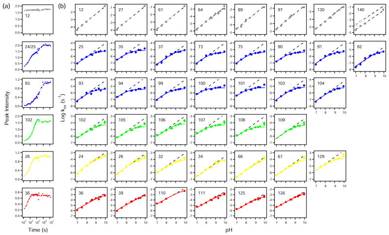
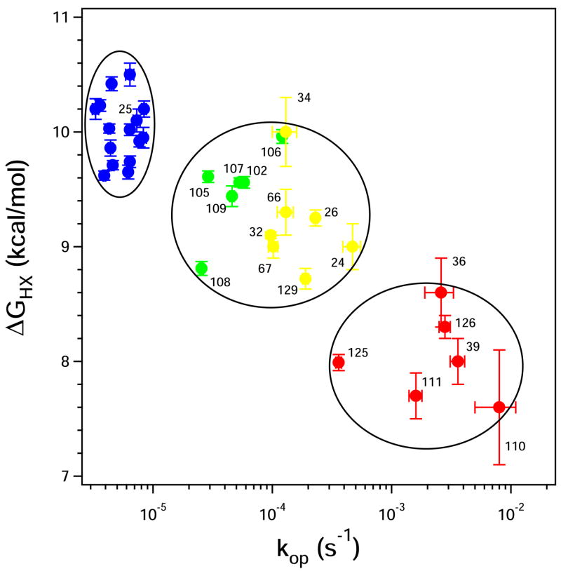
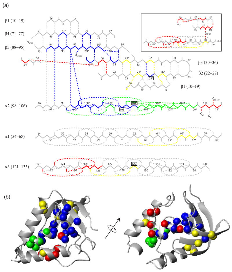
To search for submolecular foldon units, the spontaneous reversible unfolding and refolding of staphylococcal nuclease under native conditions was studied by a kinetic native-state hydrogen exchange (HX) method. As for other proteins, it appears that staphylococcal nuclease is designed as an assembly of well-integrated foldon units that may define steps in its folding pathway and may regulate some other functional properties. The HX results identify 34 amide hydrogens that exchange with solvent hydrogens under native conditions by way of large transient unfolding reactions. The HX data for each hydrogen measure the equilibrium stability (Delta G(HX)) and the kinetic unfolding and refolding rates (k(op) and k(cl)) of the unfolding reaction that exposes it to exchange. These parameters separate the 34 identified residues into three distinct HX groupings. Two correspond to clearly defined structural units in the native protein, termed the blue and red foldons. The remaining HX grouping contains residues, not well separated by their HX parameters alone, that represent two other distinct structural units in the native protein, termed the green and yellow foldons. Among these four sets, a last unfolding foldon (blue) unfolds with a rate constant of 6 x 10(-6) s(-1) and free energy equal to the protein's global stability (10.0 kcal/mol). It represents part of the beta-barrel, including mutually H-bonding residues in the beta 4 and beta 5 strands, a part of the beta 3 strand that H-bonds to beta 5, and residues at the N-terminus of the alpha2 helix that is capped by beta 5. A second foldon (green), which unfolds and refolds more rapidly and at slightly lower free energy, includes residues that define the rest of the native alpha2 helix and its C-terminal cap. A third foldon (yellow) defines the mutually H-bonded beta1-beta2-beta 3 meander, completing the native beta-barrel, plus an adjacent part of the alpha1 helix. A final foldon (red) includes residues on remaining segments that are distant in sequence but nearly adjacent in the native protein. Although the structure of the partially unfolded forms closely mimics the native organization, four residues indicate the presence of some nonnative misfolding interactions. Because the unfolding parameters of many other residues are not determined, it seems likely that the concerted foldon units are more extensive than is shown by the 34 residues actually observed.
Figures

Similar articles
-
Global analysis of the acid-induced and urea-induced unfolding of staphylococcal nuclease and two of its variants.Biochemistry. 1997 Feb 4;36(5):1129-40. doi: 10.1021/bi9609681. Biochemistry. 1997. PMID: 9033404
-
How cytochrome c folds, and why: submolecular foldon units and their stepwise sequential stabilization.J Mol Biol. 2004 Oct 8;343(1):223-33. doi: 10.1016/j.jmb.2004.08.005. J Mol Biol. 2004. PMID: 15381432
-
Fluorescence energy transfer indicates similar transient and equilibrium intermediates in staphylococcal nuclease folding.J Mol Biol. 2000 Jun 16;299(4):1133-46. doi: 10.1006/jmbi.2000.3804. J Mol Biol. 2000. PMID: 10843864
-
The nature of protein folding pathways.Proc Natl Acad Sci U S A. 2014 Nov 11;111(45):15873-80. doi: 10.1073/pnas.1411798111. Epub 2014 Oct 17. Proc Natl Acad Sci U S A. 2014. PMID: 25326421 Free PMC article. Review.
-
Protein Folding-How and Why: By Hydrogen Exchange, Fragment Separation, and Mass Spectrometry.Annu Rev Biophys. 2016 Jul 5;45:135-52. doi: 10.1146/annurev-biophys-062215-011121. Epub 2016 Apr 27. Annu Rev Biophys. 2016. PMID: 27145881 Free PMC article. Review.
Cited by
-
Structural and kinetic mapping of side-chain exposure onto the protein energy landscape.Proc Natl Acad Sci U S A. 2011 Jun 28;108(26):10532-7. doi: 10.1073/pnas.1103629108. Epub 2011 Jun 13. Proc Natl Acad Sci U S A. 2011. PMID: 21670244 Free PMC article.
-
High-Throughput Determination of Exchange Rates of Unmodified and PTM-Containing Peptides Using HX-MS.Mol Cell Proteomics. 2025 Feb;24(2):100904. doi: 10.1016/j.mcpro.2025.100904. Epub 2025 Jan 7. Mol Cell Proteomics. 2025. PMID: 39788320 Free PMC article.
-
Hydrogen Exchange Mass Spectrometry.Methods Enzymol. 2016;566:335-56. doi: 10.1016/bs.mie.2015.06.035. Epub 2015 Jul 27. Methods Enzymol. 2016. PMID: 26791986 Free PMC article.
-
Microsecond acquisition of heterogeneous structure in the folding of a TIM barrel protein.Proc Natl Acad Sci U S A. 2008 Sep 9;105(36):13367-72. doi: 10.1073/pnas.0802788105. Epub 2008 Aug 29. Proc Natl Acad Sci U S A. 2008. PMID: 18757725 Free PMC article.
-
Elucidating the mechanism of substrate recognition by the bacterial Hsp90 molecular chaperone.J Mol Biol. 2014 Jun 12;426(12):2393-404. doi: 10.1016/j.jmb.2014.04.001. Epub 2014 Apr 12. J Mol Biol. 2014. PMID: 24726919 Free PMC article.
References
-
- Bai Y, Englander SW. Future directions in folding: The multi-state nature of protein structure. Proteins: Struct Funct Genet. 1996;24:145–51. - PubMed
-
- Englander SW. Protein folding intermediates and pathways studied by hydrogen exchange. Annu Rev Biophys Biomol Struct. 2000;29:213–238. - PubMed
-
- Krishna MMG, Lin Y, Mayne L, Englander SW. Intimate view of a kinetic protein folding intermediate: Residue-resolved structure, interactions, stability, folding and unfolding rates, homogeneity. J Mol Biol. 2003;334:501–513. - PubMed
Publication types
MeSH terms
Substances
Grants and funding
LinkOut - more resources
Full Text Sources
Research Materials
Miscellaneous
