. 2008 Apr 1;228(1):93-104.
doi: 10.1016/j.taap.2007.11.032. Epub 2007 Dec 14.

The contribution of c-Jun N-terminal kinase activation and subsequent Bcl-2 phosphorylation to apoptosis induction in human B-cells is dependent on the mode of action of specific stresses

Affiliations

The contribution of c-Jun N-terminal kinase activation and subsequent Bcl-2 phosphorylation to apoptosis induction in human B-cells is dependent on the mode of action of specific stresses

Donna E Muscarella et al. Toxicol Appl Pharmacol. .

Abstract

The c-Jun N-terminal kinase (JNK) pathway can play paradoxical roles as either a pro-survival or a pro-cell death pathway depending on type of stress and cell type. The goal of the present study was to determine the role of JNK pathway signaling for regulating B-cell apoptosis in two important but contrasting situations--global proteotoxic damage, induced by arsenite and hyperthermia, versus specific microtubule inhibition, induced by the anti-cancer drug vincristine, using the EW36 B-cell line. This cell line over-expresses the Bcl-2 protein and is a useful model to identify treatments that can overcome multi-drug resistance in lymphoid cells. Exposure of EW36 B-cells to arsenite or lethal hyperthermia resulted in activation of the JNK pathway and induction of apoptosis. However, pharmacological inhibition of the JNK pathway did not inhibit apoptosis, indicating that JNK pathway activation is not required for apoptosis induction by these treatments. In contrast, vincristine treatment of EW36 B-cells resulted in JNK activation and apoptosis that was suppressed by JNK inhibition. A critical difference between the two types of stress treatments was that only vincristine-induced JNK activation resulted in phosphorylation of Bcl-2 at threonine-56, a modification that can block its anti-apoptotic function. Importantly, Bcl-2 phosphorylation was attenuated by JNK inhibition implicating JNK as the upstream kinase. Furthermore, arsenite and hyperthermia treatments activated a p53/p21 pathway associated with apoptosis induction, whereas vincristine did not activate this pathway. These results reveal two stress-activated pathways, one JNK-dependent and another JNK-independent, either of which can bypass Bcl-2 mediated resistance, resulting in cell death.


Figures
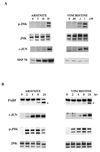
FIG 1
FIG 1
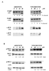
Differential activation of JNK and HSP 70-stress pathways in EW36 B-cells by arsenite compared to vincristine. (A) Protein lysates were collected from cultures exposed to arsenite or vincristine at the indicated concentrations and subjected to immunoblotting as described in Materials and Methods. The two isoforms of phosphorylated JNK (p-JNK) and total JNK protein (JNK; confirming equivalent loading of protein for each sample) were detected at 2 h following drug addition. Induction of c-Jun protein (c-JUN) and HSP 70 were determined at 24 h following drug addition. (B) Time course of JNK-pathway activation and PARP cleavage in arsenite and vincristine-treated EW36 cells. Cultures were exposed to 5 µM arsenite or 0.1 µM vincristine and protein lysates collected at the indicated times (0 h, before addition of drug up to 24 h post-drug addition). Immunoblots were performed to detect cleavage of PARP (arrow), induction of c-Jun protein and phosphorylated JNK (p-JNK). Total JNK (JNK) confirms equivalent loading of protein for each lane.
FIG 2
FIG 2
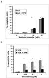
Suppression of vincristine, but not arsenite-induced apoptosis by pre-treatment with the JNK inhibitor SP6. Cultures of EW36 cells were pre-treated with DMSO control or 20 µM SP6 for 2 h prior to the addition of either arsenite (A) or vincristine (B) at the indicated concentrations. Percent of apoptotic cells was determined by staining with Hoechst 33342 and propidium iodide (H/PI) followed by diagnosis of apoptosis by fluorescence microscopy as described in Materials and Methods at 24 h. Shaded bars correspond to cultures with chemical alone, solid bars correspond to cultures treated with SP6 prior to chemical addition. Data points are from three experiments with replicate cultures and 200 cells scored per sample. A significant induction of apoptosis over control was seen in cultures exposed to arsenite at 10 and 20 µM and vincristine at all three concentrations (*; p < 0.05)). Apoptosis was significantly suppressed by SP6 pre-treatment for vincristine (**; p <0.05) but not for arsenite.
FIG 3
FIG 3
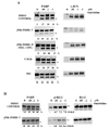
JNK-mediated phosphorylation of Bcl-2 is required for apoptosis induced by vincristine but not by arsenite. (A) Immunoblots of protein lysates collected following exposure of EW36 B-cells to arsenite or vincristine in the presence or absence of JNK inhibitor SP6 (+SP6 and −SP6, respectively) showing PARP cleavage (arrow) at 24 h and induction of c-Jun protein at 4 h following chemical addition. Percent of cleaved PARP as determined by densitometry is indicated below each lane. (B) Differential phosphorylation of Bcl-2 at thr-56. Cultures were treated with SP6 (+SP6) or DMSO control (−SP6) 2h prior to the addition of drug. Protein lysates were collected at 2 h following drug addition and subjected to immunoblotting using antibodies specific for Bcl-2 phosphorylated at thr-56 (p-Bcl-2) or total Bcl-2. The upper band corresponds to phospho-Bcl-2 and is indicated by the double asterik; the lower band is due to non-specific binding of antibody. Levels of phosphorylated Bcl-2 were determined by densitometry and are expressed as fold-increase in signal over solvent control (0 µM). Bcl-2 protein is also shown and indicates equal protein loading for all lanes.
FIG 4
FIG 4
Inhibition of vincristine-induced PARP cleavage and phosphorylation of Bcl-2 by two different JNK inhibitors, SP6 and JNK V. (A) Kinase inhibitors were added to cultures of EW36 B-cells 2 h prior to vincristine addition as follows: DMSO solvent control, JNK inhibitor V (JNK inhib.V; 10 µM); JNK inhibitor II (JNK inhib. II – negative control for SP6; 20 µM); U0126 (inhibitor of ERK; 20 µM) and SB202190 (SB2 – inhibitor of p38 kinase; 20µM). Protein lysates were made and immunoblots performed to detect PARP cleavage (arrow) at 24 h and induction of c-Jun protein at 4 h following vincristine addition. Percent of cleaved PARP as determined by densitometry is indicated below each lane. (B) Cultures were treated with 10 µM JNK inhibitor V (JNK inhib.V) or DMSO control 2 h prior to the addition of vincristine. Protein lysates were collected at 2 h following drug addition and subjected to immunoblotting using antibodies specific for Bcl-2 phosphorylated at thr-56 (p-Bcl-2) or total Bcl-2. The upper band corresponds to phospho-Bcl-2 and is indicated by the double asterik; the lower band is due to non-specific binding of antibody. Levels of phosphorylated Bcl-2 were determined by densitometry and are expressed as fold-increase in signal over solvent control (0 µM). Bcl-2 protein is also shown and indicates equal protein loading for all lanes.
FIG 5
FIG 5
Suppression of vincristine-induced apoptosis by pre-treatment with the JNK inhibitor V. Cultures of EW36 cells were pre-treated with DMSO control or 20 µM JNK inhibitor V for 2 h prior to the addition of vincristine at the indicated concentrations. Percent of apoptotic cells was determined by staining with Hoechst 33342 and propidium iodide (H/PI) followed by diagnosis of apoptosis by fluorescence microscopy as described in Materials and Methods at 24 h. Shaded bars correspond to cultures with DMSO alone, solid bars correspond to cultures treated with JNK inhibitor V (JNK V) prior to vincristine addition. Data points are from three experiments with replicate cultures and 200 cells scored per sample. A significant induction of apoptosis was seen in cultures exposed to vincristine alone at all concentrations (*; p < 0.05)). Apoptosis was significantly attenuated by JNK V pre-treatment for all three concentrations of vincristine (*; p < 0.05).
FIG 6
FIG 6
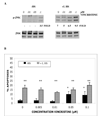
Suppression of vincristine-induced JNK and Bcl-2 phosphorylation as well as PARP cleavage by pre-treatment with non-lethal heat stress. (A) Immunoblot of heat stress protein hsp 70 (HSP 70) in protein lysates from cultures exposed to the indicated concentrations of vincristine for 24 h without prior heat stress (−HS), or following limited, non-lethal, heat stress (+NL HS; 30 min @ 43°C), or following extended, lethal heat stress, (+L HS; 1 h @ 43°C). (B) Protein lysates from cultures treated with the indicated concentrations of vincristine without prior heat stress (−HS) or subjected to limited heat stress (+NL HS) were blotted for phosphorylated Bcl-2 thr 56 (p-Bcl-2, upper band, double asterik), phosphorylated JNK (p-JNK) or total JNK protein (JNK; loading control) at 2 h following drug addition and PARP at 24 h following drug addition. Percent of PARP that is cleaved (arrow) as determined by densitometry is indicated below each lane.
FIG 7
FIG 7
Suppression of vincristine-induced apoptosis by pre-treatment with non-lethal heat stress. Percent of apoptotic cells was determined by staining with Hoechst 33342 and propidium iodide (H/PI) followed by diagnosis of apoptosis by fluorescence microscopy as described in Materials and Methods at 24 h following addition of the indicated concentrations of vincristine. Solid bars correspond to cultures with no prior treatment with heat stress (−HS), shaded bars correspond to cultures subjected to non-lethal heat stress (+NL HS) prior to drug addition. Data points are from three experiments with replicate cultures and 200 cells scored per sample. A significant induction of apoptosis was seen in cultures exposed to vincristine alone at 0.05 and 0.1 µM (*; p < 0.05)). Apoptosis was significantly suppressed in cultures exposed to non-lethal heat stress prior to vincristine addition at drug concentrations of 0.05 and 0.1 µM, (**; p < 0.05).
FIG 8
FIG 8
Effects of lethal heat stress on vincristine-induced apoptosis. (A) Protein lysates were collected from cultures treated with vincristine at the indicated concentrations without prior heat treatment (−HS) or with prior treatment with lethal heat stress (+L HS) and blotted for phosphorylated JNK (p-JNK) or total JNK (JNK; loading control) at 2 h following drug addition. (B) Percent of apoptotic cells 24 h after drug addition at the indicated concentrations determined by staining using H/PI as described in Materials and Methods. Solid bars correspond to cultures without prior treatment with heat stress (−HS), shaded bars correspond to cultures subjected to lethal heat stress (+L HS) prior to drug addition. Data points are from three experiments with replicate cultures and 200 cells scored per sample. Apoptosis was significantly increased in cultures exposed to vincristine alone starting at concentrations of 0.05 µM (*; p < 0.05) and in all cultures treated with lethal heat stress, with or without the addition of vincristine (**; p < 0.05).
FIG 9
FIG 9
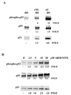
Activation of the p53 pathway and induction of the p53 target gene, p21. (A) Immunoblot of cultures not subjected to heat treatment (−HS) or exposed to non-lethal heat stress (+NL HS) or lethal heat stress (+L HS). Protein lysates were collected at 24 h following treatment and blotted for phosphorylated p53 at ser-15 (phospho-p53), total p53 protein, (p53), or p21. Fold increase in signal, as determined by densitometry, is indicated below each lane. (B) Immunblot of protein lysates from cultures exposed to indicated concentrations of arsenite 24 h after drug addition and blotted for phospho-p53, total 53 and p21 proteins as above.
FIG 10
FIG 10
Proposed pathways for JNK-dependent and JNK-independent apoptosis induction by arsenite and vincristine. (A) Vincristine induced microtubule damage leads to phosphorylation of JNK which, in turn, translocates to the nucleus and phosphorylates the transcription factor c-Jun leading to the induction of multiple nuclear substrates, including c-Jun itself. In addition, activated JNK phosphorylates the cytoplasmic substrate, Bcl-2 at thr-56 leading to its inactivation. The JNK inhibitor SP6 blocks c-Jun induction as well as Bcl-2 phosphorylation leading to an inhibition of apoptosis. Non-lethal heat stress also attenuates vincristine-induced phosphorylation of JNK and Bcl-2 thus blocking the induction of apoptosis. Whether this is due to the stabilization of microtubules by heat stress proteins or by direct effects of hypethermia on the JNK pathway is presently unknown. (B) Global proteotoxic damage induced by arsenite exposure leads to the phosphorylation of JNK that, in turn, phosphorylates the transcription factor c-Jun, leading to the induction of nuclear substrates, including c-Jun itself. However, phosphorylation of the cytoplasmic substrate Bcl-2 does not occur and inhibiting JNK with SP6 does not attenuate apoptosis. Lethal heat stress or arsenite (but not vincristine or non-lethal heat stress) induce a robust p53 response that is associated with apoptosis induction in this scenario.

Similar articles

Cited by

References

    1. Bennett BL, Sasaki DT, Murray BW, O’Leary EC, Sakata ST, Xu W, Leisten JC, Motiwala A, Pierce S, Satoh Y, Bhagwat SS, Manning AM, Anderson DW. SP600125, an anthrapyrazolone inhibitor of Jun-N-terminal kinase. Proc. Natl.Acad. Sci. (USA) 2001;98:13681–13686. - PMC - PubMed
    1. Bhalla KN. Microtubule-targeted anticancer agents and apoptosis. Oncogene. 2003;22:9075–9086. - PubMed
    1. Bloom SE, Lemley AT, Muscarella DE. Potentiation of apoptosis by heat stress plus pesticide exposure in stress resistant human B-lymphoma cells and its attenuation through interaction with follicular dendritic cells: role for c-Jun N-terminal kinase signaling. Toxicol. Sci. 2006;89:214–223. - PubMed
    1. Chen Y-R, Wang X, Templeton D, Davis RJ, Tan T-H. The role of c-Jun N-terminal kinase (JNK) in apoptosis induced by ultraviolet C and gamma radiation. J. Biol.Chem. 1996;271:31929–31936. - PubMed
    1. Deng X, Gao F, Flagg T, Anderson J, May WS. Bcl2's flexible loop domain regulates p53 binding and survival. Mol Cell Biol. 2006;26:4421–4434. - PMC - PubMed

Publication types

MeSH terms