. 2008 Mar 21;283(12):7733-44.
doi: 10.1074/jbc.M707142200. Epub 2008 Jan 16.

Amyloidogenic processing but not amyloid precursor protein (APP) intracellular C-terminal domain production requires a precisely oriented APP dimer assembled by transmembrane GXXXG motifs

Affiliations

Amyloidogenic processing but not amyloid precursor protein (APP) intracellular C-terminal domain production requires a precisely oriented APP dimer assembled by transmembrane GXXXG motifs

Pascal Kienlen-Campard et al. J Biol Chem. .

Erratum in

  • J Biol Chem. 2008 May 2;283(18):12680

Abstract

The beta-amyloid peptide (Abeta) is the major constituent of the amyloid core of senile plaques found in the brain of patients with Alzheimer disease. Abeta is produced by the sequential cleavage of the amyloid precursor protein (APP) by beta- and gamma-secretases. Cleavage of APP by gamma-secretase also generates the APP intracellular C-terminal domain (AICD) peptide, which might be involved in regulation of gene transcription. APP contains three Gly-XXX-Gly (GXXXG) motifs in its juxtamembrane and transmembrane (TM) regions. Such motifs are known to promote dimerization via close apposition of TM sequences. We demonstrate that pairwise replacement of glycines by leucines or isoleucines, but not alanines, in a GXXXG motif led to a drastic reduction of Abeta40 and Abeta42 secretion. beta-Cleavage of mutant APP was not inhibited, and reduction of Abeta secretion resulted from inhibition of gamma-cleavage. It was anticipated that decreased gamma-cleavage of mutant APP would result from inhibition of its dimerization. Surprisingly, mutations of the GXXXG motif actually enhanced dimerization of the APP C-terminal fragments, possibly via a different TM alpha-helical interface. Increased dimerization of the TM APP C-terminal domain did not affect AICD production.


Figures

Fig. 1
Fig. 1
Expression and processing of human APP GxxxG mutants in CHO cells. A. Schematic representation of the transmembrane and juxtamembrane domains of human APP695. The positions of the three consecutive GxxxG motifs (621-625, 625-629, 629-633) are highlighted. The amino acid substitution (G to A or G to L) generated for each mutant appears in bold underlined. The cleavage sites of α-(α), β- (β and β′) and γ- (γ and ε) secretase activities are indicated by arrows. The epitopes of the human-specific WO-2 antibody are also shown along with the C-terminal position recognized by the Aβ40 and Aβ42 specific antibodies for ELISA. B. The expression of cellular APP695 or APP mutants was analyzed 48 after transfection by Western blotting revealed by the WO-2 antibody. The presence of APP is indicated by arrows. Actin was used as a protein loading control. Forty hours after transfection, CHO cells were conditioned in fresh culture medium for 8 h. The accumulation of soluble APPα (sαAPP) was analyzed by Western blotting revealed by the WO-2 antibody. Aβ was immunoprecipitated from the same culture medium and analyzed by Western blotting revealed by the WO-2 antibody. C. The Aβ to sαAPP ratio was calculated and represented as percentage of Aβ/sαAPP production in non-mutated controls (APP695). Values are means ± sem, n = 4; *p < 0.05, ** p < 0.01, *** p < 0.001, compared to control.
Fig. 2
Fig. 2
GG 625/629 LL mutation does not modify the cellular distribution of APP. A. The particulate distribution of APP695 and APP mutant 5 (GG625/629LL) was analyzed in CHO cells by floatation in sucrose gradient and Western blotting revealed by the WO-2 antibody. Arrows indicate the expected positions of APP. B. Density distributions are represented as normalized histograms. C. The presence of APP at the plasma membrane was studied by cell surface biotinylation of CHO cells expressing APP and APP mutant 5. The presence of full length APP was detected by Western blotting using the human-specific WO-2 antibody in total (T), intracellular (IC) and cell surface (S) fractions. Arrows indicate the expected positions of APP. D. Quantification of surface biotinylation experiments. The cell surface APP (APPs) to total APP (APPt) ratio was calculated and given as percentage. Values are means ± sem, n = 5, ns = non-significant.
Fig. 3
Fig. 3
GG 625/629 LL mutation does not impair β-cleavage. A. Schematic representation of the APP fragments produced by β-secretase activity (sβAPP and CTFβ) along with the epitope recognized by the APP C-terminal antibody (C-ter). B. APP-BACE1 interaction was analyzed in CHO cell lines stably expressing APP695 and APP mut5 (GG625/629LL) or co-expressing APP and BACE1 by non-denaturating (Blue Native, BN) electrophoresis. Levels of BACE1 and APP in cell extracts was measured by Western blotting revealed with the WO-2 or BACE1 antibody in denaturating conditions (SDS PAGE, upper panel). Arrows indicate the expected position of cellular monomeric BACE1 or APP. The formation of cellular APP/BACE1 complexes was investigated by Blue Native electrophoresis in 6%-10% polyacrylamide gels (BN PAGE, lower panel) followed by Western blotting revealed with the WO-2 or BACE1 antibody. Arrows indicate the expected position of monomeric APP (mon APP), monomeric BACE (mon BACE1) or APP/BACE1 complexes. C. APP/BACE1 interaction analyzed by co-immunoprecipitation. Levels of BACE1 and APP were measured by Western blotting in cell extracts prior to immunoprecipitation. Cell lysates were immunoprecipitated either by the WO-2 or by the anti BACE1 antibody and revealed by Western blotting with the WO-2 or the BACE1 antibody as indicated. Arrows indicate the expected position of cellular BACE1 or APP. D. Soluble αAPP and soluble βAPP were analyzed in the same sample (extracellular medium) by multiplex assays. The production of human sβAPP from non-transfected (Co) cells and cells expressing APP695 and APP mut5 is given in ng/ml of culture medium (left) or as sβAPP/sαAPP ratio represented as percentage of APP 695. Values are means ± sem, n = 6; ns = non-significant. E. Expression of full-length APP and βCTF revealed with the C-terminal antibody.
Fig. 4
Fig. 4
Effects of GG 625/629 LL mutation on γ-cleavage and Aβ production. A. Schematic representation of the APP695 and C99 proteins. The epitope recognized by the WO-2 antibody is underlined. B. The expression of APP695, APP mut5 was measured by Western blotting in cell lysates revealed by the WO-2 antibody. The presence of APP is indicated by an arrow. C. Aβ1-40 and Aβ1-42 production was monitored by ELISA in the culture medium of cells expressing APP or APP mut5, and given as Aβ levels in pg/ml. Values are means ± sem, n = 4; * p < 0.05, *** p < 0.001, compared to control. D. The expression of C99, C99 mut5 was measured by Western blotting in cell lysates revealed by the WO-2 antibody. The presence of C99 is depicted by an arrow, the asterisk indicates the presence of possible C99 dimers. E. Aβ1-40 and Aβ1-42 production was monitored by ELISA in the culture medium of cells expressing C99 or C99 mut5, and given as Aβ levels in pg/ml. Values are means ± sem, n = 4; *** p < 0.001, compared to control.
Fig. 5
Fig. 5
GG 625/629 LL mutation does not impair APP-PS1 interaction. The interaction between APP, APP mut5 and PS1 was studied in CHO cells co-expressing APP and human PS1. A. The levels of APP (top), PS1 holoprotein and PS1 N-terminal fragment (NTF, bottom) were monitored by Western blotting revealed with the WO-2 and MAB1563 antibodies, respectively. B. Cell lysates were immunoprecipitated by the WO-2 (human APP) antibody and revealed by Western blotting with the human PS1-specific MAB1563 antibody. Arrows indicate the expected position of cellular PS1 holoprotein, and PS1 N-terminal fragment.
Fig. 6
Fig. 6
GG 625/629 LL mutation does not affect AICD release. The release of AICD by ε-cleavage of APP was studied in CHO cells expressing either APP695 or APPGal4 fusion proteins. A. Schematic representation of the APPGal4 protein in comparison to APP695, along with the epitope recognized by the WO-2 and APP C-terminal antibody (C-ter). B. Expression of APP695, APPGal4 and APPGal4 mut5 was measured in transfected CHO cells by Western blotting revealed by the WO-2 antibody. C. Aβ1-40 production was monitored by ELISA in the culture medium of cells expressing APPGal4, APPGal4 mut5, and given as Aβ levels in pg/ml, nd = non-detectable (below 15 pg/ml). Values are means ± sem, n = 4; *** p < 0.001, compared to control. D. The release of AICD was measured by a Gal4 transactivation assay (bottom). Luciferase activity was normalized and represented as activity relative to control (APP695). Values are means ± sem, n = 4, ns = non-significant, *** p < 0.001, compared to control. E. Forty hours after transfection, CHO cells were treated for 8 h by 100μM of 1-10 Phenanthroline monohydrate (orthophenantroline, PNT), a metalloprotease inhibitor. AICD levels were measured in cell lysates (Western blotting) revealed by the C-ter antibody. Arrows indicate the expected position of αCTF and AICD. F. The same experiments were performed in cells expressing APP695 or APP mut5. AICD levels were measured in cell lysates (Western blotting) revealed by the C-ter antibody. Arrows indicate the expected position of APP, αCTF and AICD.
Fig. 7
Fig. 7
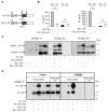
GG 625/629 LL mutation triggers the formation of C99 homodimers. A. Schematic representation of the HA- and Myc-tagged C99 proteins. The epitope recognized by the WO-2 antibody is underlined. B. Aβ1-40 and Aβ1-42 production was monitored by ELISA in the culture medium of cells expressing HA-C99, Myc-C99, or the corresponding mutants 5, and given as Aβ levels in pg/ml. Values are means ± sem, n = 4; *** p < 0.001, compared to control. C. The expression of HA- and Myc-C99 and HA- and Myc-C99 mut5 was measured by Western blotting in cell lysates revealed by the anti-HA (left), anti-Myc (middle) and WO-2 (right) antibodies. The presence of tagged C99 at the expected molecular weight is indicated by an arrow. The asterisk indicates the presence of possible C99 dimers. D. The formation of C99 homodimers was analyzed in CHO cell lines stably expressing HA-C99, Myc-C99 or the corresponding mutants 5. Levels of C99 was measured by Western blotting revealed by the anti-HA antibody in cell extracts prior to immunoprecipitation (Input, direct lysates). Extracts from cells expressing separately the HA-C99 and Myc-C99 were mixed and analyzed by Western blotting prior to immunoprecipitation (Input, post-lysate mix). Cell lysates were further immunoprecipitated by the anti-Myc and revealed by Western blotting with the anti-HA antibody as indicated. Arrows indicate the expected position of HA-C99 monomers (HA-C99) and HA-C99 dimers (HA-C99*).
Fig. 8
Fig. 8
G to L and G to I mutations display similar effects on Aβ production, AICD release and C99 oligomers formation. A. Schematic representation of the transmembrane (TM) and juxtamembrane domains of the C99 G to L or G to I mutants. B. The expression of C99 and C99 mutants was measured by Western blotting in cell lysates revealed by the WO-2 antibody (Top). The presence of C99 is depicted by an arrow, the asterisk indicates the presence of C99 SDS-resistant dimers. The presence of AICD and αCTFs was measured in the same cell lysates by Wetern blotting revealed with the C-ter antibody (Bottom). D. Aβ1-40 and Aβ1-42 production was monitored by ELISA in the culture medium of cells expressing the indicated G to L or G to I C99 mutants. Results are given as Aβ levels in pg/ml. Values are means ± sem, n = 4; *** p < 0.001, compared to control.
Fig. 9
Fig. 9
Dimer models for the wild-type APP transmembrane domain, the GG 625/629 LL and the G 629 I mutants. A. Low energy dimer of the TM domain of APP. The dimer interface is lined by glycines at positions 621, 625, 629 and 633. B-C. Low energy dimer of the TM domain of the GG 625/629 LL and G 629 I mutants of APP, respectively. Mutation of glycines 625 and 629 to leucine, or glycine 629 to isoleucine results in rotation of the transmembrane helices. C. Helix-helix interaction energies for the dimer structures for wild-type APP (solid line, full circles), the GG 625/629 LL mutant (dashed line, open circles) and G 629 I mutant (dashed line, open squares). The glycines in the interface of wild-type APP dimer allow Ser622 to form a strong interhelical hydrogen bond. In the GG/LL and G/I mutants, the Gly634xxxAla638 sequence allows close approach of the helices. In these mutants, interhelical hydrogen bonding of Asn623 provides the most stabilizing interaction.

Similar articles

Cited by

References

    1. Seubert P, Vigo-Pelfrey C, Esch F, Lee M, Dovey H, Davis D, Sinha S, Schlossmacher M, Whaley J, Swindlehurst C. Nature. 1992;359:325–327. - PubMed
    1. Shoji M, Golde TE, Ghiso J, Cheung TT, Estus S, Shaffer LM, Cai XD, McKay DM, Tintner R, Frangione B. Science. 1992;258:126–129. - PubMed
    1. Kang J, Lemaire HG, Unterbeck A, Salbaum JM, Masters CL, Grzeschik KH, Multhaup G, Beyreuther K, Muller-Hill B. Nature. 1987;325:733–736. - PubMed
    1. Sinha S, Anderson JP, Barbour R, Basi GS, Caccavello R, Davis D, Doan M, Dovey HF, Frigon N, Hong J, Jacobson-Croak K, Jewett N, Keim P, Knops J, Lieberburg I, Power M, Tan H, Tatsuno G, Tung J, Schenk D, Seubert P, Suomensaari SM, Wang S, Walker D, John V. Nature. 1999;402:537–540. - PubMed
    1. Vassar R, Bennett BD, Babu-Khan S, Kahn S, Mendiaz EA, Denis P, Teplow DB, Ross S, Amarante P, Loeloff R, Luo Y, Fisher S, Fuller J, Edenson S, Lile J, Jarosinski MA, Biere AL, Curran E, Burgess T, Louis JC, Collins F, Treanor J, Rogers G, Citron M. Science. 1999;286:735–741. - PubMed

Publication types

Substances

LinkOut - more resources