. 2008 Jan;7(1):79-89.
doi: 10.1158/1535-7163.MCT-07-0584.

Modulation of the antitumor activity of metronomic cyclophosphamide by the angiogenesis inhibitor axitinib

Affiliations

Modulation of the antitumor activity of metronomic cyclophosphamide by the angiogenesis inhibitor axitinib

Jie Ma et al. Mol Cancer Ther. 2008 Jan.

Abstract

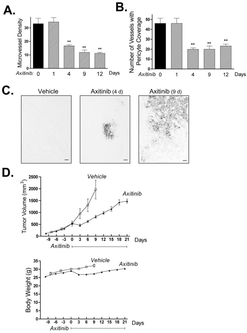
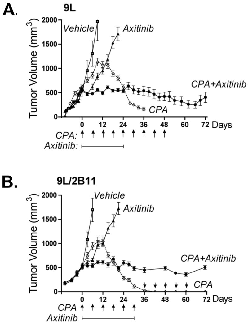
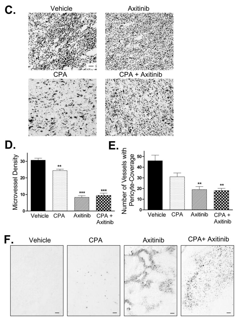
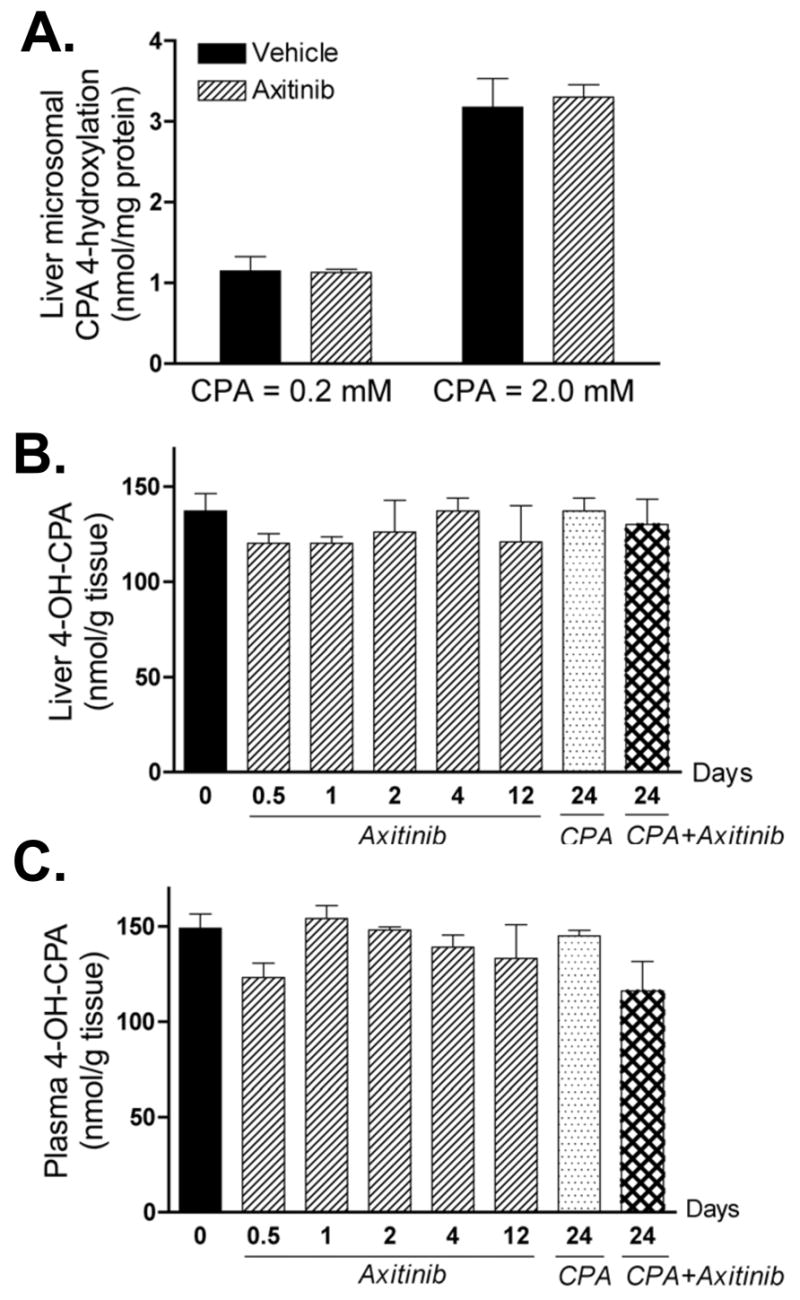
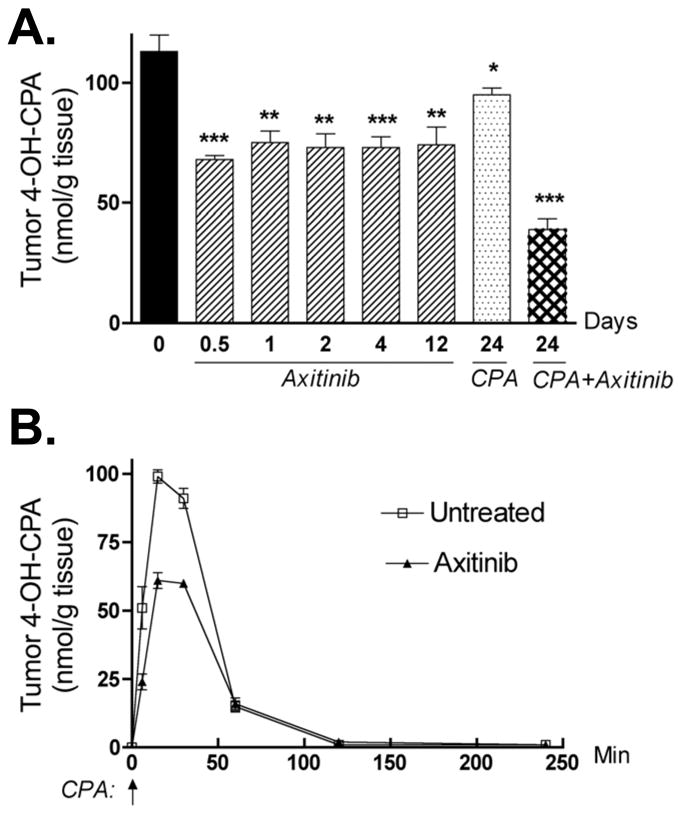
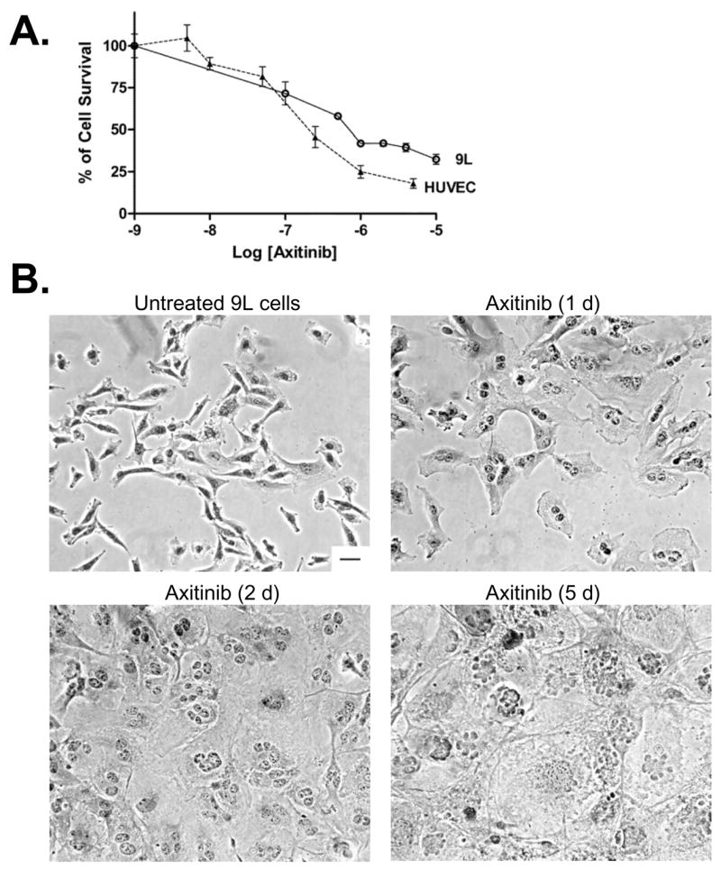
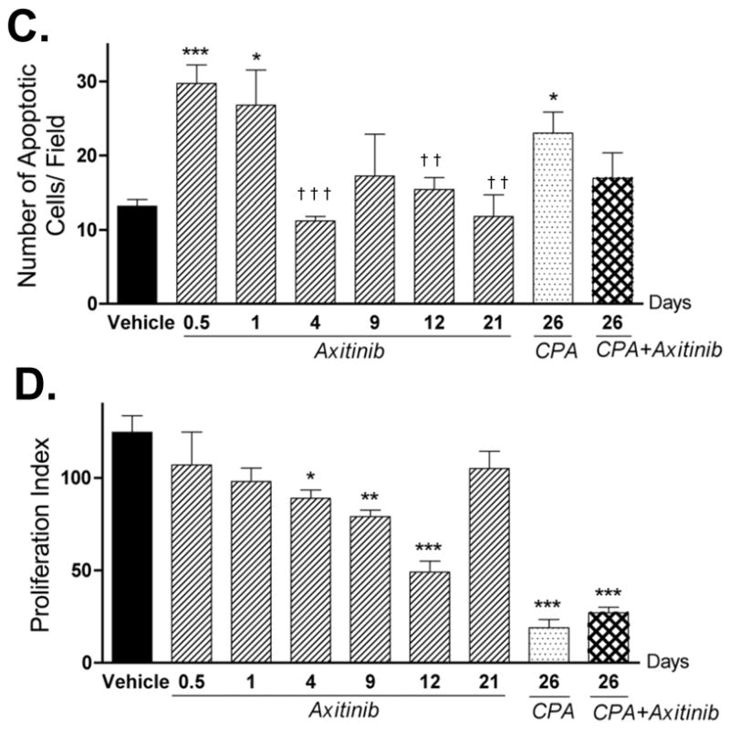
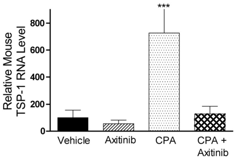
The promising but still limited efficacy of angiogenesis inhibitors as monotherapies for cancer treatment indicates a need to integrate these agents into existing therapeutic regimens. Presently, we investigate the antitumor activity of the small-molecule angiogenesis inhibitor axitinib (AG-013736) and its potential for combination with metronomic cyclophosphamide. Axitinib significantly inhibited angiogenesis in rat 9L tumors grown s.c. in scid mice but only moderately delayed tumor growth. Combination of axitinib with metronomic cyclophosphamide fully blocked 9L tumor growth on initiation of drug treatment. In contrast, metronomic cyclophosphamide alone required multiple treatment cycles to halt tumor growth. However, in contrast to the substantial tumor regression that is ultimately induced by metronomic cyclophosphamide, the axitinib/cyclophosphamide combination was tumor growth static. Axitinib did not inhibit hepatic activation of cyclophosphamide or export of its activated metabolite, 4-hydroxy-cyclophosphamide (4-OH-CPA), to extrahepatic tissues; rather, axitinib selectively decreased 9L tumor uptake of 4-OH-CPA by 30% to 40%. The reduced tumor penetration of 4-OH-CPA was associated with a decrease in cyclophosphamide-induced tumor cell apoptosis and a block in the induction of the endogenous angiogenesis inhibitor thrombospondin-1 in tumor-associated host cells, which may contribute to the absence of tumor regression with the axitinib/cyclophosphamide combination. Finally, axitinib transiently increased 9L tumor cell apoptosis, indicating that its effects are not limited to the endothelial cell population. These findings highlight the multiple effects that may characterize antiangiogenic agent/metronomic chemotherapy combinations and suggest that careful optimization of drug scheduling and dosages will be required to maximize antitumor responses.


Figures

Figure 1
Figure 1. Anti-angiogenic activity and tumor growth delay of axitinib monotherapy in rat 9L gliosarcoma
9L tumors grown s.c. in scid mice were treated with axitinib for up to 21 days at 25 mg/kg, i.p., sid, as specified in each panel. A. Number of tumor blood vessels per CD31-immunostained 9L tumor section counted at 400x magnification. Axitinib significantly reduced microvessel density after 4–12 days of treatment. B. Impact of axitinib treatment on the number of 9L tumor blood vessels with pericyte coverage, identified as SMA-α-positive blood vessels at 200x magnification. Days of axitinib treatment are indicated along the x-axis, with n = 4 tumors/group and ** p < 0.01 compared to day 0 controls (panels A and B). C. Immunostaining for hypoxia-specific dye pimonidazole revealed an increase in tumor hypoxia after 4 and 9 days of axitinib treatment. Scale bar at bottom right, 100 μm. D. Axitinib treatment initiated on day 0 delayed 9L tumor growth (upper panel, n = 10 tumors/group) with minimal effect on the rate of mouse body weight gain (lower panel). Solid line along x-axis indicates the time period of daily axitinib treatment.
Figure 2
Figure 2. Axitinib/metronomic CPA combination blocks 9L tumor growth but does not induce tumor regression
A. and B. 9L tumors (panel A) or 9L/2B11 tumors, which can activate CPA intratumorally (panel B), were implanted s.c. in scid mice, grown to an average vol of 500 mm3 and then treated with the axitinib/metronomic CPA combination (see Materials and Methods). CPA-induced tumor regression (most complete for 9L/2B11 tumors) was absent in the combination treatment group, which displayed sustained tumor growth stasis, continuing even after termination of drug treatment. Arrows along the x-axis indicate days of CPA treatment (140 mg/kg, i.p., every 6-d) while solid line below the arrows indicates the time period of axitinib treatment (25 mg/kg, i.p., sid). On the days when both drugs were co-administered, CPA was given 4 hr prior to axitinib to minimize drug-drug interactions. For 9L tumors (A), n = 20 to 24 tumors/group through day 24, after which 6 tumors were left for longer-term monitoring while the other tumor samples were collected for analysis. In case of 9L/2B11 tumors (panel C), n = 10 tumors/group through day 30, after which both drug treatments were terminated for the CPA + axitinib combination group and 3 mice (6 tumors) were removed for tissue analysis; the remaining 4 tumors were monitored till day 72. For the CPA-treated 9L/2B11 tumors, CPA administration on an every 6-day schedule was continued for the 4 remaining tumors from day 30 through day 60 (arrows above x-axis) and tumor measurements continued till day 72. C. 9L tumor cryosections prepared from mice receiving the indicated treatments were stained with hematoxylin-eosin. Similar tumor cell morphologies and cell densities were observed in tumors from mice treated with vehicle, axitinib (25 mg/kg, i.p., sid, 21 days), or the combination therapy (26 days). In contrast, metronomic CPA (140 mg/kg, i.p., every 6-d for 26 days) induced changes in both cell morphology and density. Scale bar at bottom right of vehicle-treated tumors panel applies to all four graphs, 50 μm. D. Microvessel density of CD31-immunostained 9L tumor cryosections measured at 400x magnification. E. Number of blood vessels with SMA-α-positive pericyte coverage counted at 200x magnification. F. Anti-angiogenic activity of the axitinib/CPA combination was manifest by increased tumor hypoxia as detected by pimonidazole staining. Scale bar, 100 μm. Drug treatments for panels D, E and F were the same as described in panel C. Panels D and E, n =4 tumors/group, **, p<0.01 and ***, p<0.001 compared to vehicle controls.
Figure 3
Figure 3. Impact of axitinib treatment on hepatic activation of CPA and export of its active metabolite, 4-OH-CPA
A. Mice bearing 9L tumors were treated with vehicle or axitinib (25 mg/kg, i.p. sid) for 4 days. 24 hr after the last drug injection, liver microsomes were isolated (21) and CPA 4-hydroxylase activity was assayed by HPLC (23) following in vitro incubation with 0.2 or 2 mM of CPA. Data shown are mean ± SE based on n = 3 livers/group. The levels of 4-OH-CPA in liver (B) and plasma (C) were measured by HPLC 15 min after injection of a single test dose of CPA (140 mg/kg, i.p.). The 15 min time point corresponds to the Tmax (4-OH-CPA) (see Fig. 4B, below). None of the values shown is significantly different than the day 0 (untreated) controls (n = 3 mice/group, p > 0.05 for all treatments compared to controls). Numbers along the x-axis indicate the time period of drug treatment, as described in Fig. 2.
Figure 4
Figure 4. Impact of axitinib or axitinib/CPA combination on 9L tumor 4-OH-CPA exposure
A. Levels of 4-OH-CPA in 9L tumors treated with axitinib (25 mg/kg, i.p. sid), metronomic CPA (140 mg/kg, i.p., every 6-d) or the combination for the indicated periods of time were measured 15 min after injection of a test dose of CPA, as described in Fig. 3. Intratumoral 4-OH-CPA uptake was decreased ~ 40% within 12 hr after the first axitinib injection and remained at that level through 12 daily axitinib treatments. Four cycles of the combination treatment decreased 4-OH-CPA uptake by 9L tumors >60%. * p < 0.05, ** p < 0.01, and *** p < 0.001 compared to day 0 controls, with n = 6 tumors/group. B. Levels of 4-OH-CPA in untreated or axitinib-pretreated (25 mg/kg, i.p., sid for 4 d) 9L tumors were measured 6 to 240 min after a single test dose of CPA (single i.p. injection at 140 mg/kg, indicated by the vertical arrow at 0 min). AUC values for tumor 4-OH-CPA exposure decreased from 4545 to 3251 nmol/g*min after 4 d of axitinib treatment (see Table S1), with n = 6 tumors/time point.
Figure 5
Figure 5. Direct effects of axitinib on 9L tumor cells
A. Cultured 9L tumor cells and HUVEC cells were treated with axitinib at the indicated concentrations for 4 d and the impact on final cell number, indicative of relative growth rate, was determined by crystal violet staining (n = 3 replicates/data point). B. Morphology of hematoxylin-eosin stained 9L cells treated with axitinib (500 nM) for up to 5 d in culture. Scale bar at bottom right of untreated cells panel applies to all graphs, 50 μm. The impact of the indicated in vivo drug treatments on tumor cell apoptosis (C) and proliferation (D) was analyzed by TUNEL assay and PCNA immunostaining, respectively. The number of staining-positive cells was counted at 200x magnification. Numbers along the x-axis indicate the time period of drug treatment. * p < 0.05, ** p < 0.01, and *** p < 0.001 compared to day 0 controls, while † p < 0.05, †† p < 0.01, and ††† p < 0.001 compared to the response at 12 hr (day 0.5), n = 4 tumors/group.
Figure 6
Figure 6. Expression of mouse TSP-1 RNA after multiple drug treatments
The level of host (mouse) TSP-1 RNA in 9L tumor was measured by real-time PCR following treatment with axitinib (25 mg/kg, i.p., sid for 21 d), metronomic CPA (140 mg/kg, i.p., every 6-d for 26 d) or the combination (26 d). Metronomic CPA induced a 7-fold increase in mouse TSP-1 expression, which was absent in axitinib or the combination therapy-treated tumors. Data are expressed as relative RNA levels, mean ± SE for n = 4–8 individual tumors/group.

Similar articles

Cited by

References

    1. Jain RK, Duda DG, Clark JW, Loeffler JS. Lessons from phase III clinical trials on anti-VEGF therapy for cancer. Nat Clin Pract Oncol. 2006;3(1):24–40. - PubMed
    1. Ferrara N, Kerbel RS. Angiogenesis as a therapeutic target. Nature. 2005;438(7070):967–74. - PubMed
    1. Ellis LM. Mechanisms of action of bevacizumab as a component of therapy for metastatic colorectal cancer. Semin Oncol. 2006;33(5 Suppl 10):S1–7. - PubMed
    1. Kerbel RS. Antiangiogenic therapy: a universal chemosensitization strategy for cancer? Science. 2006;312(5777):1171–5. - PubMed
    1. Nieder C, Wiedenmann N, Andratschke N, Molls M. Current status of angiogenesis inhibitors combined with radiation therapy. Cancer Treat Rev. 2006;32(5):348–64. - PubMed

Publication types

MeSH terms