

Automatic classification of MR scans in Alzheimer's disease
- PMID: 18202106
- PMCID: PMC2579744
- DOI: 10.1093/brain/awm319
Automatic classification of MR scans in Alzheimer's disease
Abstract
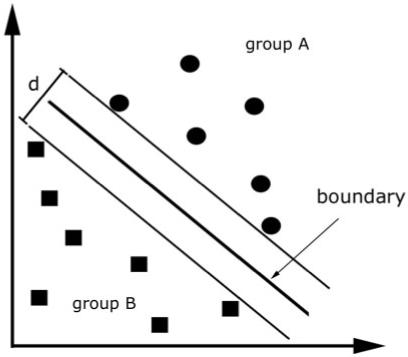
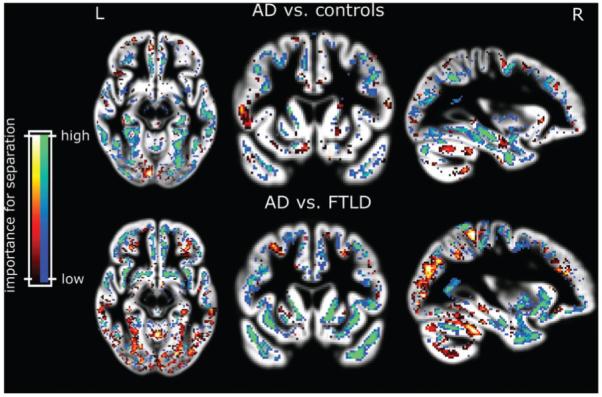
To be diagnostically useful, structural MRI must reliably distinguish Alzheimer's disease (AD) from normal aging in individual scans. Recent advances in statistical learning theory have led to the application of support vector machines to MRI for detection of a variety of disease states. The aims of this study were to assess how successfully support vector machines assigned individual diagnoses and to determine whether data-sets combined from multiple scanners and different centres could be used to obtain effective classification of scans. We used linear support vector machines to classify the grey matter segment of T1-weighted MR scans from pathologically proven AD patients and cognitively normal elderly individuals obtained from two centres with different scanning equipment. Because the clinical diagnosis of mild AD is difficult we also tested the ability of support vector machines to differentiate control scans from patients without post-mortem confirmation. Finally we sought to use these methods to differentiate scans between patients suffering from AD from those with frontotemporal lobar degeneration. Up to 96% of pathologically verified AD patients were correctly classified using whole brain images. Data from different centres were successfully combined achieving comparable results from the separate analyses. Importantly, data from one centre could be used to train a support vector machine to accurately differentiate AD and normal ageing scans obtained from another centre with different subjects and different scanner equipment. Patients with mild, clinically probable AD and age/sex matched controls were correctly separated in 89% of cases which is compatible with published diagnosis rates in the best clinical centres. This method correctly assigned 89% of patients with post-mortem confirmed diagnosis of either AD or frontotemporal lobar degeneration to their respective group. Our study leads to three conclusions: Firstly, support vector machines successfully separate patients with AD from healthy aging subjects. Secondly, they perform well in the differential diagnosis of two different forms of dementia. Thirdly, the method is robust and can be generalized across different centres. This suggests an important role for computer based diagnostic image analysis for clinical practice.
Figures
Comment in
-
Reply: A plea for confidence intervals and consideration of generalizability in diagnostic studies.Brain. 2009 Apr;132(Pt 4):e103; author reply e102. doi: 10.1093/brain/awn090. Brain. 2009. PMID: 19382289 Free PMC article. No abstract available.
References
-
- American Psychiatric Association . Diagnostic, and statistical manual of mental disorders. American Psychiatric Press; Washington, DC: 1987.
-
- Ashburner J. A fast diffeomorphic image registration algorithm. Neuroimage. 2007;38:95–113. - PubMed
-
- Ashburner J, Friston KJ. Voxel-based morphometry—the methods. Neuroimage. 2000;11:805–21. - PubMed
-
- Barnes J, Scahill RI, Boyes RG, Frost C, Lewis EB, Rossor CL, et al. Differentiating AD from aging using semiautomated measurement of hippocampal atrophy rates. Neuroimage. 2004;23:574–81. - PubMed
-
- Bishop C. Pattern recognition and machine learning. Springer; New York: 2006.
Publication types
MeSH terms
Grants and funding
LinkOut - more resources
Full Text Sources
Other Literature Sources
Medical
