. 2008 May;149(5):2108-20.
doi: 10.1210/en.2007-0582. Epub 2008 Jan 17.

Biochemical and structural characterization of apolipoprotein A-I binding protein, a novel phosphoprotein with a potential role in sperm capacitation

Affiliations

Biochemical and structural characterization of apolipoprotein A-I binding protein, a novel phosphoprotein with a potential role in sperm capacitation

Kula N Jha et al. Endocrinology. 2008 May.

Abstract

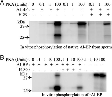
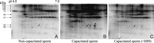
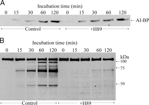
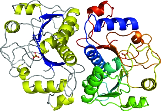
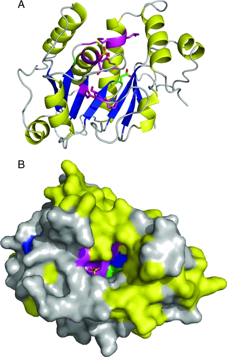
The physiological changes that sperm undergo in the female reproductive tract rendering them fertilization-competent constitute the phenomenon of capacitation. Cholesterol efflux from the sperm surface and protein kinase A (PKA)-dependent phosphorylation play major regulatory roles in capacitation, but the link between these two phenomena is unknown. We report that apolipoprotein A-I binding protein (AI-BP) is phosphorylated downstream to PKA activation, localizes to both sperm head and tail domains, and is released from the sperm into the media during in vitro capacitation. AI-BP interacts with apolipoprotein A-I, the component of high-density lipoprotein involved in cholesterol transport. The crystal structure demonstrates that the subunit of the AI-BP homodimer has a Rossmann-like fold. The protein surface has a large two compartment cavity lined with conserved residues. This cavity is likely to constitute an active site, suggesting that AI-BP functions as an enzyme. The presence of AI-BP in sperm, its phosphorylation by PKA, and its release during capacitation suggest that AI-BP plays an important role in capacitation possibly providing a link between protein phosphorylation and cholesterol efflux.


Figures

Figure 1
Figure 1
A, Autoradiograms of two-dimensional gels after in vitro phosphorylation of mouse sperm lysates with γ32P ATP in either the absence (left panel) or presence (right panel) of exogenous dbcAMP and IBMX demonstrating increase in signals of several proteins that are phosphorylated downstream to PKA activation. B, Sequence alignment of mouse AI-BP and its homologs with known crystal structures, YNL200C from S. cerevisiae (PDB: 1JZT) and tm0922 protein from T. maritima (PDB: 2AX3). The invariant and conserved residues among 387 eukaryotic, bacterial, and archaeal homologs present in Pfam database are colored in green and magenta, respectively. The secondary structure of AI-BP is shown with the corresponding conventional segments of the Rossmann-fold labeled in parentheses. The signal peptide and disordered N-terminal segment present only in mammalian homologs are in gray and black, respectively. Two peptides that identified AI-BP in mass spectrometric analysis are shown in boxes. The figure was drawn with ALSCRIPT (69).
Figure 2
Figure 2
Expression of recombinant AI-BP. A, Coomassie blue stain of lysed bacterial proteins from uninduced (U) and induced (I) cultures containing recombinant AI-BP expression constructs. B, Western blot of uninduced (U) and induced (I) bacterial protein lysates using anti-nickel NTA antibody, demonstrating recombinant AI-BP at the expected molecular mass of 29 kDa only in induced culture.
Figure 3
Figure 3
A, Western blots showing reactivity of AI-BP antiserum from the guinea pig with the recombinant AI-BP (rAI-BP) and native AI-BP from mouse sperm and testis. Preimmune serum did not react with either recombinant AI-BP or native AI-BP from mouse sperm or testis. B, Two-dimensional Western blot of mouse sperm lysate showing reactivity of AI-BP from the sperm to AI-BP antiserum from the guinea pig. Total protein loading is shown by a companion silver-stained gel that also marks the corresponding spot of AI-BP (arrow), which was cored out for the mass spectrometric analysis.
Figure 4
Figure 4
A, Autoradiogram of SDS-PAGE gel from in vitro phosphorylation of native AI-BP immunoprecipitated from mouse sperm by purified PKA catalytic subunits either in the presence or in the absence of a PKA inhibitor H89. The presence or absence of a component in the phosphorylation reaction is represented by + and − signs, respectively. B, Autoradiogram of SDS-PAGE gel from in vitro phosphorylation of recombinant AI-BP by purified PKA catalytic subunits in either the presence or absence of H89.
Figure 5
Figure 5
Autoradiograms of two-dimensional gels from in vivo phosphorylation in intact mouse sperm with [32P] orthophosphate in noncapacitation medium (A), capacitation medium (B), and capacitation medium containing H89 (C). AI-BP spot is marked by an arrow in the autoradiograms. On the top pI range and on the left, molecular masses are marked.
Figure 6
Figure 6
Mouse multiple tissue Northern blot probed with 32P-labeled cDNA of mouse AI-BP demonstrating an approximately 1.0 kb band in several tissues with highest levels of expression in heart, liver, kidney, and testis. After probing with AI-BP, the same blot was stripped and reprobed with 32P-labeled β-actin cDNA probe as a loading control (lower panel).
Figure 7
Figure 7
Immunofluorescent localization of AI-BP in mouse sperm using antiserum raised in guinea pig (A) and the preimmune serum (C). The corresponding phase-contrast micrographs of the identical fields are shown next to the respective images (B and D).
Figure 8
Figure 8
A, Western blot of AI-BP released into the capacitation medium over different time periods during capacitation in the absence (control) and presence of H89. B, Western blot of the sperm lysates probed with antiphosphotyrosine antibody in the absence (control) and presence of H89.
Figure 9
Figure 9
The AI-BP homodimer is comprised of subunits with a Rossmann-like fold. One subunit is colored according to the secondary structure (strands in blue, helices in yellow, loops in gray) and the second subunit is colored in rainbow pattern from blue N terminus to red C terminus of the polypeptide chain. The first 24 residues of mature AI-BP are disordered in the crystal structure. The sulfate anions present in crystallization media are bound above the C-terminal end of the β-sheets. All pictures representing the structure were drawn with PyMOL (30).
Figure 10
Figure 10
The putative active site of AI-BP marked by conserved residues. The AI-BP subunit is colored according to the secondary structure. The two invariant (green) and 11 highly conserved (magenta) residues (Fig. 1B) are clustered around the two compartment cavity that contains bound sulfate (stick representation). A, Ribbon view of AI-BP subunit with side chains of the invariant and highly conserved residues. B, Surface view of the subunit rotated approximately 90° around the x-axis. The trench compartment of the cavity runs along the surface from left to right and extends into the perpendicular pocket on the upper right side.

References

    1. Austin CR 1952 The capacitation of the mammalian sperm. Nature 170:326 - PubMed
    1. Yanagimachi R 1994 Mammalian fertilization. 2nd ed. New York: Raven Press
    1. Travis AJ, Kopf GS 2002 The role of cholesterol efflux in regulating the fertilization potential of mammalian spermatozoa. J Clin Invest 110:731–736 - PMC - PubMed
    1. Visconti PE, Westbrook VA, Chertihin O, Demarco I, Sleight S, Diekman AB 2002 Novel signaling pathways involved in sperm acquisition of fertilizing capacity. J Reprod Immunol 53:133–150 - PubMed
    1. Jha KN, Kameshwari DB, Shivaji S 2003 Role of signaling pathways in regulating the capacitation of mammalian spermatozoa. Cell Mol Biol 49:329–340 - PubMed

Publication types

MeSH terms