. 2008 Jan 29;105(4):1285-90.
doi: 10.1073/pnas.0711293105. Epub 2008 Jan 17.

MHC class I chain-related protein A antibodies and shedding are associated with the progression of multiple myeloma

Affiliations

MHC class I chain-related protein A antibodies and shedding are associated with the progression of multiple myeloma

Masahisa Jinushi et al. Proc Natl Acad Sci U S A. .

Erratum in

Abstract

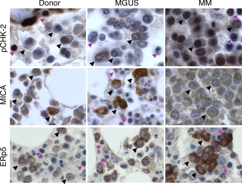
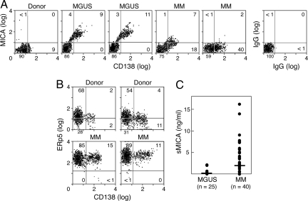
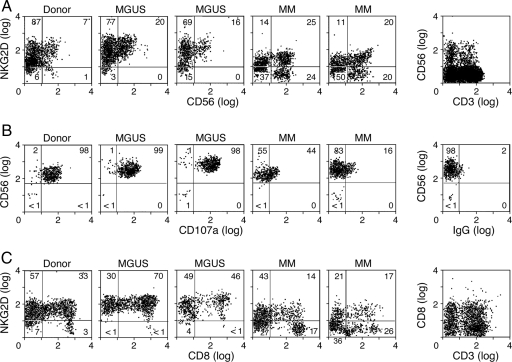
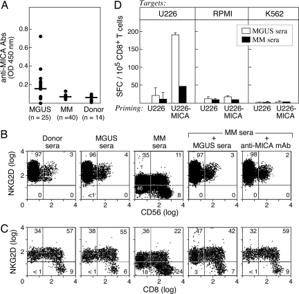
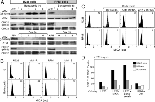
Monoclonal gammopathy of undetermined significance (MGUS) is a common disorder of aging and a precursor lesion to full-blown multiple myeloma (MM). The mechanisms underlying the progression from MGUS to MM are incompletely understood but include the suppression of innate and adaptive antitumor immunity. Here, we demonstrate that NKG2D, an activating receptor on natural killer (NK) cells, CD8(+) T lymphocytes, and MHC class I chain-related protein A (MICA), an NKG2D ligand induced in malignant plasma cells through DNA damage, contribute to the pathogenesis of MGUS and MM. MICA expression is increased on plasma cells from MGUS patients compared with normal donors, whereas MM patients display intermediate MICA levels and a high expression of ERp5, a protein disulfide isomerase linked to MICA shedding (sMICA). MM, but not MGUS, patients harbor circulating sMICA, which triggers the down-regulation of NKG2D and impaired lymphocyte cytotoxicity. In contrast, MGUS, but not MM, patients generate high-titer anti-MICA antibodies that antagonize the suppressive effects of sMICA and stimulate dendritic cell cross-presentation of malignant plasma cells. Bortezomib, a proteasome inhibitor with anti-MM clinical efficacy, activates the DNA damage response to augment MICA expression in some MM cells, thereby enhancing their opsonization by anti-MICA antibodies. Together, these findings reveal that the alterations in the NKG2D pathway are associated with the progression from MGUS to MM and raise the possibility that anti-MICA monoclonal antibodies might prove therapeutic for these disorders.


Conflict of interest statement

The authors declare no conflict of interest.

Figures

Fig. 1.
Fig. 1.
The DNA damage response is activated, and MICA and ERp5 expression are increased during MGUS/MM progression. Bone marrow tissue microarrays were prepared from healthy donors (n = 10) and patients with untreated MGUS (n = 20) or MM (n = 40) and were stained for phospho-chk-2 (Top), MICA (Middle), or ERp5 (Bottom). Representative examples from the arrays are shown. (×250 magnification.) Black arrows, plasma cells; purple arrows, nonplasma cells.
Fig. 2.
Fig. 2.
MICA and ERp5 plasma cell surface expression and MICA shedding during the progression of MM. (A) Bone marrow aspirates were stained for CD138 and MICA and analyzed with flow cytometry. The results are representative of three normal donors, three MGUS patients, and seven MM patients studied. (B) Bone marrow aspirates were stained for CD138 and ERp5. The results are representative of three normal donors and six MM patients studied. (C) Sera from MGUS and MM patients were evaluated for sMICA with an ELISA. The lower limits of the assay were 90 pg/ml.
Fig. 3.
Fig. 3.
NKG2D expression and cytotoxic lymphocyte function during the progression of MM. (A) PBMCs from normal donors and patients with MGUS or MM were evaluated for NKG2D expression on CD56+ cells by flow cytometry. The results are representative of three normal donors, three MGUS patients, and seven MM patients studied. (B) Magnetic bead-purified healthy donor, MGUS patient, or MM patient NK cells were tested for lytic activity against K562 targets by surface CD107a mobilization. The results are representative of three normal donors, three MGUS patients, and three MM patients studied. (C) PBMCs from normal donors and patients with MGUS or MM were evaluated for NKG2D expression on CD8+ cells by flow cytometry. The results are representative of three normal donors, three MGUS patients, and seven MM patients studied.
Fig. 4.
Fig. 4.
MGUS patients develop anti-MICA antibodies with functional activity. (A) Sera from MGUS or MM patients and healthy donors were diluted 1:100 and analyzed by ELISA with recombinant MICA protein. The reactivity was determined with a pan-IgG secondary. (B) Donor PBMCs were incubated for 48 h in the indicated sera, and then the NKG2D expression on CD56+ cells was determined with flow cytometry. Anti-MICA monoclonal antibodies were added where indicated. (C) CD8+ cells are shown. Similar results were observed by using three different MGUS and MM sera samples. (D) Dendritic cells were generated from adherent donor PBMCs, loaded with MGUS or MM sera-coated U226 or U226-MICA MM cells, matured with LPS, and used to stimulate autologous-purified CD8+ T cells for 7 days. IFN-γ production was measured by ELISPOT against K562 cells or dendritic cells loaded with U226 or RPMI-8226 MM cells. No reactivity against unpulsed dendritic cells or dendritic cells loaded with unopsonized tumors was observed (data not shown).
Fig. 5.
Fig. 5.
Bortezomib activates the DNA damage response and increases MICA expression in some MM cells. (A) U226 and RPMI-8226 MM cells were treated with 20 μM Bortezomib, 250 μg/ml dexamethasone, or 10 μg/ml aphidicolin (a DNA polymerase inhibitor that serves as a control for inducing DNA damage) for the indicated times. Cell lysates were prepared and evaluated for phospho-ATM (ser 1981), ATM, phospho-chk-2 (thr 68), or chk-2 by immunoblotting. (B) The indicated MM cells were treated with 20 μM Bortezomib overnight, and surface MICA expression was determined with flow cytometry. (C) U226 and RPMI-8226 MM cells were transfected with plasmids encoding control, ATM, or chk-2 shRNAs; exposed to Bortezomib overnight; and assayed for MICA expression with flow cytometry. (D) Dendritic cells were generated from adherent donor PBMCs; loaded with MGUS, MM, or normal sera-coated U226, U226-MICA, Bortezomib-treated U226, or dexamethasone-treated U226 MM cells; matured with LPS; and used to stimulate autologous-purified CD8+ T cells for 7 days. IFN-γ production was measured by ELISPOT against dendritic cells loaded with U226 MM cells. No reactivity against K562 cells, unpulsed dendritic cells, or dendritic cells loaded with unopsonized tumors was observed (data not shown). Results are representative of four donors. Points were performed in triplicate. Means are shown, with SD < 10%.

References

    1. Kyle RA, et al. Prevalence of monoclonal gammopathy of undetermined significance. N Engl J Med. 2006;354:1362–1369. - PubMed
    1. Kyle RA, et al. A long-term study of prognosis in monoclonal gammopathy of undetermined significance. N Engl J Med. 2002;346:564–569. - PubMed
    1. International Myeloma Working Group. Criteria for the classification of monoclonal gammopathies, multiple myeloma and related disorders: A report of the International Myeloma Working Group. Br J Haematol. 2003;121:749–757. - PubMed
    1. Davies FE, et al. Insights into the multistep transformation of MGUS to myeloma using microarray expression analysis. Blood. 2003;102:4504–4511. - PubMed
    1. Kuehl WM, Bergsagel PL. Multiple myeloma: Evolving genetic events and host interactions. Nat Rev Cancer. 2002;2:175–187. - PubMed

Publication types

MeSH terms