. 2008 Jan 29;105(4):1221-5.
doi: 10.1073/pnas.0710338104. Epub 2008 Jan 17.

Plant cyclotides disrupt epithelial cells in the midgut of lepidopteran larvae

Affiliations

Plant cyclotides disrupt epithelial cells in the midgut of lepidopteran larvae

Barbara L Barbeta et al. Proc Natl Acad Sci U S A. .

Abstract

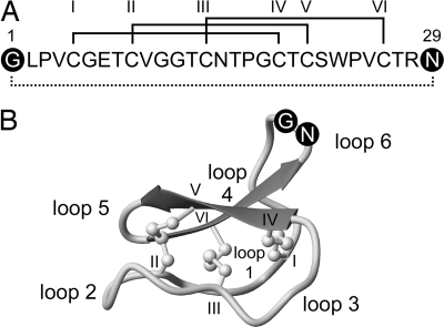
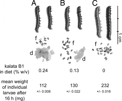
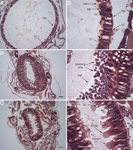
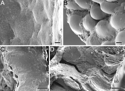
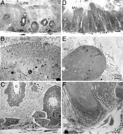
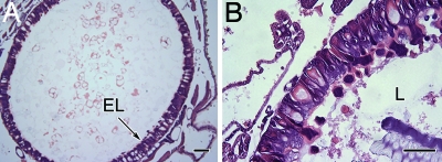
Several members of the Rubiaceae and Violaceae plant families produce a series of cyclotides or macrocyclic peptides of 28-37 aa with an embedded cystine knot. The cyclic peptide backbone together with the knotted and strongly braced structure confers exceptional chemical and biological stability that has attracted attention for potential pharmaceutical applications. Cyclotides display a diverse range of biological activities, such as uterotonic action, anti-HIV activity, and neurotensin antagonism. In plants, their primary role is probably protection from insect attack. Ingestion of the cyclotide kalata B1 severely retards the growth of larvae from the Lepidopteran species Helicoverpa armigera. We examined the gut of these larvae after consumption of kalata B1 by light, scanning, and transmission electron microscopy. We established that kalata B1 induces disruption of the microvilli, blebbing, swelling, and ultimately rupture of the cells of the gut epithelium. The histology of this response is similar to the response of H. armigera larvae to the Bacillus thuringiensis delta-endotoxin, which is widely used to control these insect pests of crops such as cotton.


Conflict of interest statement

The authors declare no conflict of interest.

Figures

Fig. 1.
Fig. 1.
Sequence and structure of kalata B1. (A) Amino acid sequence of kalata B1 showing disulfide connectivities (roman numerals) and the circular peptide backbone (dotted line). The first and last residues in the mature domain are in black circles (Gly-1 and Asn-29). (B) A ribbon structure of mature kalata B1 (Protein Data Bank ID code 1NB1) (39). The six cysteine residues are labeled with roman numerals, and the six intercysteine segments (loops) are numbered 1–6. Loop 6 is completed by the formation of a new peptide bond between Gly-1 and Asn-29 to produce the circular peptide backbone. The acyclic kalata B1 permutant has a break in the peptide bond between Gly-1 and Asn-29.
Fig. 2.
Fig. 2.
The appearance and mean average weight of third-instar larvae after 16 h on control diet (C) or diets containing 0.24% (wt/vol) (A) or 0.13% (wt/vol) (B) kalata B1. Diet (d) not consumed after 16 h and the amount of fecal pellets (f) produced by an individual larvae from each group are also shown. As the concentration of kalata B1 increased less diet was consumed and fewer fecal pellets were produced.
Fig. 3.
Fig. 3.
Light microscopy of the anterior midgut third-instar larvae fed for 16 h on control and kalata B1 diets. Columnar cells (CC), goblet cells (GC), brush border (BB) of the columnar cells, lumen (L), peritrophic membrane (PM), and epithelial layer (EL) are as labeled. Stem cells (SC) are located along the basement membrane (BM) between columnar and goblet cells. (A and D) The midgut epithelium of third-instar larva after ingestion of control diet (16 h). (B and E) Kalata B1-fed [0.13% (wt/vol)] larva. The diameter of the gut is smaller than the gut in control larvae. The epithelial layer appears thicker because of blebbing of epithelial cells into the gut lumen. (C and F) Kalata B1-fed [0.24% (wt/vol)] larva. The width of the cell layer was much greater than the width of that in the control larvae. CCs were elongated and swollen and some had burst releasing granular material into the lumen. The loss of brush border at the apex of columnar cells is indicated by arrows. The peritrophic membrane was not visible. At the basement membrane the number of stems cells had increased significantly. (Scale bars: A–D, 100 μm; E and F, 50 μm.)
Fig. 4.
Fig. 4.
Scanning electron micrographs of anterior midgut epithelia after ingestion of control (A and C) and kalata B1 [0.24% (wt/vol)] (B and D) diets. The luminal surface is shown in A and B, and a transverse section from freeze fracture is shown in C and D. Gut from kalata B1-fed larvae had columnar cells that were swollen and protruding into the lumen and granular material (GM) was entangled with the peritrophic membrane (D). Brush border (BB), basement membrane (BM), a swollen cell (labeled 1), a nonswollen columnar cell (labeled 2), and peritrophic membrane (PM) are indicated. (Scale bars: 10 μm.)
Fig. 5.
Fig. 5.
Transmission electron microscopy of the anterior midgut epithelia of midsecond-instar larvae fed control diet (A–C) and diet supplemented with kalata B1 [0.24% (wt/vol)] (D–F). A and D show a cross section of the complete epithelium comprising columnar cells (C) and goblet cells (G). B and E show the apical regions of columnar cells with apical microvilli (MV), mitochondria (M), lipid droplets (L), and glycogen deposits (GL). The peritrophic membrane is evident in control larvae (B). C and F show the basal region of columnar cells with arrows indicating basal infoldings. (Scale bars: 2 μm.)
Fig. 6.
Fig. 6.
The effects of linear kalata B1 protein on the midgut of third-instar larvae after 16 h. (A) Control tissue. (B) Linear kalata B1 [0.24% (wt/vol)] produced increased shedding of cells from the epithelial layer (EL) into the lumen (L). No other damage was observed. (Scale bars: A, 100 μm; B, 50 μm.)

References

    1. Craik DJ, Daly NL, Bond T, Waine C. J Mol Biol. 1999;294:1327–1336. - PubMed
    1. Craik DJ, Cemazar M, Wang CK, Daly NL. Biopolymers. 2006;84:250–266. - PubMed
    1. Saether O, et al. Biochemistry. 1995;34:4147–4158. - PubMed
    1. Colgrave ML, Craik DJ. Biochemistry. 2004;43:5965–5975. - PubMed
    1. Craik DJ. Science. 2006;311:1563–1564. - PubMed

Publication types

MeSH terms

LinkOut - more resources