. 2008 Feb;172(2):395-405.
doi: 10.2353/ajpath.2008.070870. Epub 2008 Jan 17.

Galectin-3 reduces the severity of pneumococcal pneumonia by augmenting neutrophil function

Affiliations

Galectin-3 reduces the severity of pneumococcal pneumonia by augmenting neutrophil function

Sarah L Farnworth et al. Am J Pathol. 2008 Feb.

Abstract

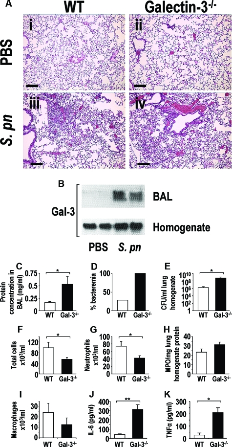
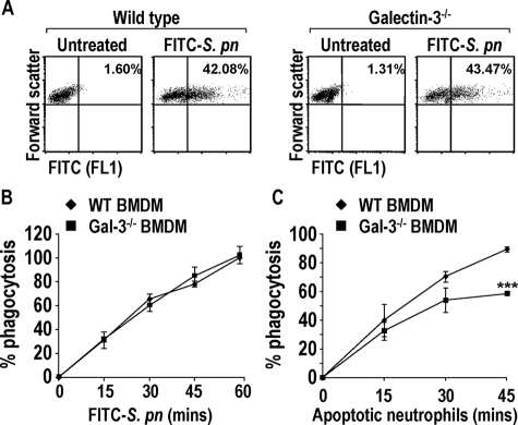
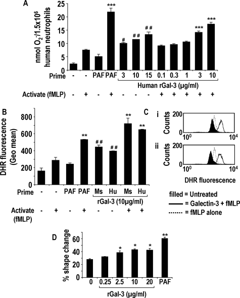
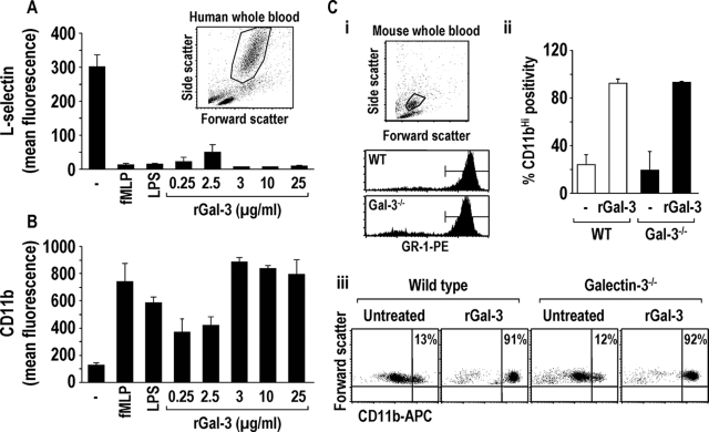
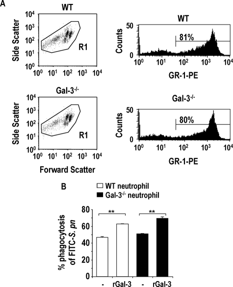
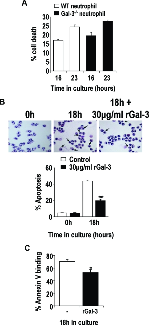
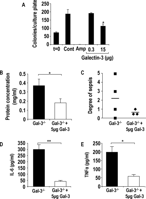
The Gram-positive Streptococcus pneumoniae is the leading cause of community-acquired pneumonia worldwide, resulting in high mortality. Our in vivo studies show that galectin-3(-/-) mice develop more severe pneumonia after infection with S. pneumoniae, as demonstrated by increased bacteremia and lung damage compared to wild-type mice and that galectin-3 reduces the severity of pneumococcal pneumonia in part by augmenting neutrophil function. Specifically, we show that 1) galectin-3 directly acts as a neutrophil-activating agent and potentiates the effect of fMLP, 2) exogenous galectin-3 augments neutrophil phagocytosis of bacteria and delays neutrophil apoptosis, 3) phagocytosis of apoptotic neutrophils by galectin-3(-/-) macrophages is less efficient compared to wild type, and 4) galectin-3 demonstrates bacteriostatic properties against S. pneumoniae in vitro. Furthermore, ad-back of recombinant galectin-3 in vivo protects galectin-3-deficient mice from developing severe pneumonia. Together, these results demonstrate that galectin-3 is a key molecule in the host defense against pneumococcal infection. Therapeutic strategies designed to augment galectin-3 activity may both enhance inflammatory cell function (by directly affecting neutrophil responsiveness and prolonging neutrophil longevity) and have direct bacteriostatic activity, improving clinical outcomes after severe pneumococcal infection.


Figures

Figure 1
Figure 1
Galectin-3 plays a critical role in the clearance of an acute pneumococcal infection. Mice were inoculated intratracheally with 1 × 10⁵ CFU S. pneumoniae for 15 hours (n = 10 mice in each group). A: H&E staining of WT and galectin-3−/− lung tissue from control (i and ii) (PBS instilled intratracheally) and S. pneumoniae pneumonia (iii and iv) (S. pneumoniae inoculated intratracheally) at 15 hours after instillation. B: Representative galectin-3 Western blots showing galectin-3 levels in BAL (neat) and homogenates (1:10) of WT mice inoculated with S. pneumoniae compared to PBS control. C–K: WT (white bars) and galectin-3−/− (black bars). C: Protein concentration was significantly higher in lavage fluid from galectin-3−/− mice compared with WT (*P < 0.05 compared to WT). D: Blood from galectin-3−/− and WT mice was plated on blood agar plates and the percentage of plates with bacterial growth is expressed as percent bacteremia. E: Lungs from galectin-3−/− and WT mice were homogenized, serially diluted, plated on blood agar plates, and bacterial counts determined. Galectin-3−/− lung homogenate demonstrated higher bacterial counts compared to WT (*P < 0.05 compared to WT). Total cell recruitment (F) and neutrophil recruitment (G) into the alveolar space was reduced in galectin-3−/− mice compared to WT mice after S. pneumoniae infection (*P < 0.05 compared to WT). H: Myeloperoxidase activity in lung homogenate was similar between the two groups. I: Macrophage recruitment into the alveolar space of WT and galectin-3−/− mice was similar. IL-6 (J) and TNF-α (K) concentration in BAL was determined by CBA and demonstrated to be higher in galectin-3-deficient animals (**P < 0.01 compared to WT and **P < 0.05 compared to WT). Scale bars = 100 μm.
Figure 2
Figure 2
Expression of galectin-3 in macrophages does not enhance phagocytosis of S. pneumoniae but endogenous galectin-3 augments macrophage phagocytosis of apoptotic human neutrophils. A: Representative forward scatter versus FL1 dot blots of WT and galectin-3−/− BMDMs treated for 60 minutes with 10:1 ratio of FITC-S. pneumoniae. Gated on macrophages according to forward and side scatter properties. Percent in upper right quadrant indicates macrophages that have phagocytosed FITC-labeled bacteria. B: Phagocytosis of a 10:1 ratio of opsonized FITC-S. pneumoniae for up to 1 hour by WT (♦) and galectin-3−/− (▪) BMDMs measured using FACS analysis. Results represent the mean percentage phagocytosis (compared to WT 60 minutes) (n = 3). C: Phagocytosis of a 10:1 ratio of apoptotic human neutrophils for up to 45 minutes by WT (♦) and galectin-3−/− (▪) BMDMs measured using FACS analysis. Results represent the mean percent phagocytosis (n = 3) (***P < 0.0001 compared to WT 45 minutes).
Figure 3
Figure 3
Exogenously added recombinant galectin-3 causes human neutrophil activation and the generation of ROS. Free radical generation from human neutrophils treated with PAF (1 μmol/L) or human galectin-3 (0.1 to 15 μg/ml) for 15 minutes followed by fMLP (0.1 μmol/L) or PBS for 15 minutes was measured by cytochrome c reduction (A) (n = 6) (***P < 0.0001 compared to treatment with fMLP alone, #P < 0.05 and ##P < 0.01 compared to treatment with PAF alone) and DHR fluorescence using FACS analysis (B and C) (n = 3) (**P < 0.01 compared to treatment with fMLP alone and ##P < 0.01 compared to treatment with PAF alone). C: Representative histogram showing DHR fluorescence in response to fMLP pretreated with either 10 μg/ml mouse (i) or human recombinant galectin-3 (ii). D: Percentage of shape change of human neutrophils treated with galectin-3 (0.25 to 20 μg/ml) or 1 μmol/L PAF for 15 minutes (n = 3) (*P < 0.05 and **P < 0.01 compared with untreated).
Figure 4
Figure 4
Exogenously added recombinant galectin-3 causes l-selectin shedding and CD11b up-regulation on human whole blood neutrophils and CD11b up-regulation on both WT and galectin-3−/− mouse neutrophils. l-selectin shedding (A) and CD11b up-regulation (B) on human neutrophils after a 30-minute incubation with fMLP (0.1 μmol/L), lipopolysaccharide (100 ng/ml), or human galectin-3 (0.25 to 25 μg/ml) was assessed by FACS analysis (n = 6) (for all treatments P < 0.01 compared to untreated). A, inset: Representative dot blot showing forward and side scatter properties of human whole blood, gated on neutrophils. C: i: Representative dot blot showing forward and side scatter properties of WT mouse whole blood gated on GR-1-positive neutrophils. ii and iii: Mouse whole blood was treated with 30 μg/ml of mouse galectin-3 for 30 minutes and incubated with anti-CD11b-APC and anti-GR-1-PE. Galectin-3-treated WT (white bars) and galectin-3−/− (black bars) whole blood demonstrated increased CD11b expression (CD11bHI) on GR-1-positive neutrophils as measured by FACS analysis (n = 3).
Figure 5
Figure 5
Exogenous galectin-3 enhances phagocytosis of S. pneumoniae by both WT and galectin-3−/− neutrophils. A: Representative dot blots from WT and galectin-3−/− mouse bone marrow neutrophil preparations gated on all populations (R1). Representative histograms demonstrate ∼80% GR-1 positivity within the R1 gate. B: WT (white bars) and galectin-3−/− (black bars) bone marrow neutrophils were prepared as described in the Materials and Methods. Neutrophils were incubated with 30 μg/ml of recombinant mouse galectin-3 or PBS (control) for 1 hour at 37°C then incubated with a 10:1 ratio of opsonized FITC-S. pneumoniae for 1 hour at 37°C. Samples were gated on GR-1-positive cells and analyzed using a BD FACSCalibur (n = 3) (**P < 0.01 compared to untreated).
Figure 6
Figure 6
WT and galectin-3−/− mouse neutrophils undergo apoptosis at similar rates in culture, and galectin-3 protects human neutrophils from apoptosis. A: Mouse bone marrow neutrophils were cultured for the indicated time points and incubated with PE-conjugated anti-GR-1 antibody followed by annexin-V-FITC. ToPro-3 was added to each sample before FACS analysis. Apoptotic and necrotic neutrophils were identified as those demonstrating GR-1 and annexin-V/ToPro-3 positivity (n = 3). B: Human peripheral blood neutrophils were cultured for 18 hours in Iscove’s modified Dulbecco’s medium containing 10% FBS and 30 μg/ml of human recombinant galectin-3 or PBS. Cytospin preparations were prepared, and neutrophil apoptosis was determined by morphology. Arrows show apoptotic human neutrophils. Percent apoptosis was calculated from total cell number of five fields of each slide (n = 3) (**P < 0.01 compared to untreated). C: Human peripheral blood neutrophils were cultured for 18 hours in Iscove’s modified Dulbecco’s medium containing 10% FBS and 20 μg/ml of human recombinant galectin-3. Neutrophils were incubated with annexin-V-FITC, and ToPro-3 was added to each sample before FACS analysis. Annexin-V-positive and ToPro-3-negative neutrophils were classed as apoptotic (n = 3) (*P < 0.05 compared to untreated).
Figure 7
Figure 7
Galectin-3 is bacteriostatic, and delivery of recombinant galectin-3 into the lungs of galectin-3−/− mice reduces severity of pneumonia. A: S. pneumoniae cultures were incubated with galectin-3 (0.3 and 15 μg) or ampicillin (20 μg/ml) for 2 hours, diluted 1:10, and plated onto blood agar plates. Colonies were counted the following day (n = 3) (*P < 0.05 compared to control). Galectin-3-deficient mice were intratracheally inoculated with 1 ×10⁵ CFU S. pneumoniae in the presence or absence of 5 μg of recombinant mouse galectin-3 for 15 hours. B: Protein concentration was significantly higher in lavage fluid from galectin-3−/− (black bars) mice compared with galectin-3−/− mice treated with galectin-3 (white bars) (*P < 0.05 compared to galectin-3−/−). C: Blood from galectin-3−/− and WT mice was plated on blood agar plates, and the degree of bacterial presence per plate was scored according to the arbitrary scale 0 to 5 (0, no bacterial growth; 5, significant bacterial growth). Galectin-3−/− mice treated with galectin-3 demonstrated reduced sepsis compared to untreated galectin-3−/− mice. IL-6 (D) and TNF-α (E) concentration in BAL was determined by CBA and demonstrated to be lower in galectin-3-treated galectin-3−/− mice (**P < 0.01 compared to galectin-3−/− and *P < 0.05 compared to galectin-3−/−).

Similar articles

Cited by

References

    1. Paterson GK, Blue CE, Mitchell TJ. Role of interleukin-18 in experimental infections with Streptococcus pneumoniae. J Med Microbiol. 2005;54:323–326. - PubMed
    1. Feldman C. Clinical relevance of antimicrobial resistance in the management of pneumococcal community-acquired pneumonia. J Lab Clin Med. 2004;143:269–283. - PubMed
    1. Novak R, Henriques B, Charpentier E, Normark S, Tuomanen E. Emergence of vancomycin tolerance in Streptococcus pneumoniae. Nature. 1999;399:590–593. - PubMed
    1. Sibille Y, Reynolds HY. Macrophages and polymorphonuclear neutrophils in lung defense and injury. Am Rev Respir Dis. 1990;141:471–501. - PubMed
    1. Barondes SH, Cooper DN, Gitt MA, Leffler H. Galectins. Structure and function of a large family of animal lectins. J Biol Chem. 1994;269:20807–20810. - PubMed

Publication types