. 2008 Feb;172(2):545-53.
doi: 10.2353/ajpath.2008.070938. Epub 2008 Jan 17.

The galectin profile of the endothelium: altered expression and localization in activated and tumor endothelial cells

Affiliations

The galectin profile of the endothelium: altered expression and localization in activated and tumor endothelial cells

Victor L Thijssen et al. Am J Pathol. 2008 Feb.

Abstract

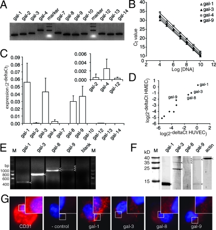
We previously identified overexpression of galectin-1 in activated tumor endothelium. Currently, the tumor vasculature is a target for therapeutic approaches. Little is known about galectin expression and regulation in the tumor vasculature. Here, we report the expression of galectin-1/-3/-8/-9 in the endothelium as determined by quantitative PCR, Western blot, flow cytometry, and immunohistochemistry. Galectin-2/-4/-12 were detectable at the mRNA level, albeit very low. Galectin-8 and -9 displayed alternative splicing. Immunohistochemistry of normal tissues revealed a broad but low expression of galectin-1 in the vasculature, whereas the expression levels and localization of the other galectins varied. Endothelial cell activation in vitro significantly increased the expression of galectin-1 (5.32 +/- 1.97; P = 0.04) and decreased the expression of both galectin-8 (0.59 +/- 0.12; P < 0.04) and galectin-9 (0.32 +/- 0.06; P < 0.002). Galectin-3 expression was unaltered. Although a portion of these proteins is expressed intracellularly, the membrane protein level of galectin-1/-8/-9 was significantly increased on cell activation in vitro, 6-fold (P = 0.005), 3-fold (P = 0.002), and 1.4-fold (P = 0.04), respectively. Altered expression levels and cellular localization was also observed in vivo in the endothelium of human tumor tissue compared with normal tissue. These data show that endothelial cells express several members of the galectin family and that their expression and distribution changes on cell activation, resulting in a different profile in the tumor vasculature. This offers opportunities to develop therapeutic strategies that are independent of tumor type.


Figures

Figure 1
Figure 1
Galectin expression profile in endothelial cells. A: Agarose gel electrophoresis showing the length of the different galectin PCR amplicons. All primers generated a single band of the expected size. B: Analysis of primer sensitivity. The graph shows linear amplification with a slope of ∼−3.3 over a broad dilution range. Only galectin-1, -3, -4, and -9 are shown, but similar results were obtained for all galectins (see also Table 3). C: The galectin expression profile in freshly isolated HUVECs. Apart from the predominantly expressed galectin-1, -3, -8, and -9, expression was also detected for galectin-2, -4, and -12 (see inset). D: Scatterplot showing a strong correlation (r = 0.967, P < 0.001) between galectin expression in HUVECs (x axis) and human microvascular endothelial cells (y axis). Galectin-1, -3, -8, and -9 were expressed most abundantly. E: Agarose gel electrophoresis following PCR with full-length primers on HUVEC cDNA. Different amplicons are indicated with an arrowhead. As blank no primers were added to the PCR mix. F: Western blot analysis of galectin protein expression in HUVECs. Protein bands are indicated with an arrowhead. Actin served as control. G: Fluorescent microscopic pictures of galectin staining (red) in HUVECs after cytospin. As positive control, a CD31 antibody was used. In the negative control, the primary antibody was omitted. Cells were counterstained with 4′,6-diamidino-2-phenylindole to visualize the nucleus (blue). Original magnification, ×1000.
Figure 2
Figure 2
Galectin expression in the endothelium of different human tissues. Immunohistochemical staining of galectin expression in different human tissues, ie, placenta (A, E, I, M, and Q), liver (B, F, J, N, and R), kidney (C, G, K, O, and S), and colon (D, H, L, P, and T). As positive vessel staining, CD31/34 was used (A–D, brown staining). Staining was performed for galectin-1 (E–H), galectin-3 (I–L), galectin-8 (M–P), and galectin-9 (Q–T). Each inset shows the endothelial cells in more detail with asterisks marking the luminal site of the vessel. The lower inset in A–D shows the negative control in which the primary antibody was omitted.
Figure 3
Figure 3
Galectin expression and localization after endothelial cell activation. A: Fold change in mRNA expression, measured by qPCR, of galectins in cultured HUVECs (black bars) compared with native HUVECs (white bars, set to 1). B: Effect of additional activation of cultured cells (black bars, set to 1) with tumor conditioned culture medium (gray bars) on mRNA expression, measured by qPCR. C: Analysis of total galectin protein levels (white bars) and extracellular, membrane-bound galectin protein (black bars) in native HUVEC measured by FACS. D: Effect of cell activation on total galectin protein expression levels. E: Effect of cell activation on extracellular, membrane-bound galectin protein levels. *P < 0.05 versus native cells. F: Relative increase in extracellular versus total galectin protein levels in response to cell activation. *P < 0.05 versus native cells.
Figure 4
Figure 4
Galectin expression in activated tumor endothelial cells. Immunohistochemical staining of galectin expression in human colon carcinoma tissue. Staining was performed for CD31/34 (A), galectin-1 (B), galectin-3 (C), galectin-8 (D), and galectin-9 (E). As negative control, the primary antibody was omitted (F). Bar = 50 um. Each inset shows a vessel in more detail with asterisks marking the luminal site of the vessel. The black arrows point toward positive stained endothelial cells. In F, the arrows point toward nuclei of unstained endothelial cells. Bar = 10 μm.

References

    1. Liu FT, Rabinovich GA. Galectins as modulators of tumour progression. Nat Rev Cancer. 2005;5:29–41. - PubMed
    1. van den Brûle F, Califice S, Castronovo V. Expression of galectins in cancer: a critical review. Glycoconj J. 2004;19:537–542. - PubMed
    1. Huflejt ME, Leffler H. Galectin-4 in normal tissues and cancer. Glycoconj J. 2004;20:247–255. - PubMed
    1. Bidon-Wagner N, Le Pennec JP. Human galectin-8 isoforms and cancer. Glycoconj J. 2004;19:557–563. - PubMed
    1. Griffioen AW, Molema G. Angiogenesis: potentials for pharmacologic intervention in the treatment of cancer, cardiovascular diseases, and chronic inflammation. Pharmacol Rev. 2000;52:237–268. - PubMed