Mechanisms of occlusion and recanalization in canine carotid bifurcation aneurysms embolized with platinum coils: an alternative concept
- PMID: 18202238
- PMCID: PMC7978218
- DOI: 10.3174/ajnr.A0902
Mechanisms of occlusion and recanalization in canine carotid bifurcation aneurysms embolized with platinum coils: an alternative concept
Abstract
Background and purpose: Endovascular treatment of aneurysms may result in complete or incomplete occlusions or may be followed by recurrences. The goal of the present study was to better define pathologic features associated with so-called healing or recurrences after coiling and to propose an alternative concept to the currently accepted view.
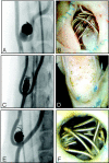
Materials and methods: Experimental canine venous pouch aneurysms were created by using a T-type (group A, N = 29) or a Y-type constructed bifurcation (group B, N = 37) between the carotid arteries. Coil embolization was performed 2 weeks later; and angiography, immediately after and at 12 weeks. Angiographic results, neointima formation at the neck, endothelialization, and organization of thrombus were compared between groups by using qualitative scores and immunohistochemistry.
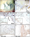
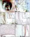
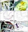
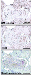
Results: Angiographic results at 3 months were significantly better in group A than in group B (P = .001). Macroscopic neointimal scores were also better (P = .012). Only 10/32 aneurysms with satisfactory results at angiography were completely sealed by neointima formation. Animals with residual or recurrent aneurysms had significantly worse neointimal scores than those with completely occluded ones (P = .0003). On histologic sections, the neointima was constantly present in "healed" and in recurrent aneurysms. This neointima was a multicellular layer of alpha-actin+ cells in a collagenous matrix, covered with a single layer of nitric oxide synthetase (NOS+) endothelial cells, whether it completely occluded the neck of the aneurysm or dived into the recurring or residual space between the aneurysm wall and the coil mass embedded in organizing thrombus.
Conclusion: Complete angiographic occlusions at 3 months can be associated with incomplete neointimal closure of the neck at pathology. Thrombus organization, endothelialization, and neointima formation can occur concurrently with recurrences.
Figures



Similar articles
-
Matrix metalloproteinase-9 may play a role in recanalization and recurrence after therapeutic embolization of aneurysms or arteries.J Vasc Interv Radiol. 2007 Oct;18(10):1271-9. doi: 10.1016/j.jvir.2007.06.034. J Vasc Interv Radiol. 2007. PMID: 17911518
-
In vivo embolization of lateral wall aneurysms in canines using the liquid-to-solid gelling PPODA-QT polymer system: 6-month pilot study.J Neurosurg. 2013 Jul;119(1):228-38. doi: 10.3171/2013.3.JNS121865. Epub 2013 Apr 5. J Neurosurg. 2013. PMID: 23560578
-
Role of the endothelial lining in recurrences after coil embolization: prevention of recanalization by endothelial denudation.Stroke. 2004 Jun;35(6):1471-5. doi: 10.1161/01.STR.0000126042.76153.f7. Epub 2004 Apr 22. Stroke. 2004. PMID: 15105520
-
Embolization with autologous fibroblast-attached platinum coils in canine carotid artery aneurysms: histopathological differences from plain coil embolization.Invest Radiol. 2003 May;38(5):281-7. doi: 10.1097/01.RLI.0000064698.12359.79. Invest Radiol. 2003. PMID: 12750617
-
Endovascular treatment of experimental wide neck aneurysms: comparison of results using coils or cyanoacrylate with the assistance of an aneurysm neck bridge device.AJNR Am J Neuroradiol. 2002 Nov-Dec;23(10):1710-6. AJNR Am J Neuroradiol. 2002. PMID: 12427629 Free PMC article.
Cited by
-
Evaluation of an endovascular aneurysm model in pigs for chronic experiments.Brain Circ. 2025 Mar 21;11(1):77-85. doi: 10.4103/bc.bc_112_24. eCollection 2025 Jan-Mar. Brain Circ. 2025. PMID: 40224549 Free PMC article.
-
Creation of large elastase-induced aneurysms: presurgical arterial remodeling using arteriovenous fistulas.AJNR Am J Neuroradiol. 2010 Nov;31(10):1935-7. doi: 10.3174/ajnr.A2205. Epub 2010 Jul 15. AJNR Am J Neuroradiol. 2010. PMID: 20634302 Free PMC article.
-
Sitagliptin Stimulates Endothelial Progenitor Cells to Induce Endothelialization in Aneurysm Necks Through the SDF-1/CXCR4/NRF2 Signaling Pathway.Front Endocrinol (Lausanne). 2019 Nov 26;10:823. doi: 10.3389/fendo.2019.00823. eCollection 2019. Front Endocrinol (Lausanne). 2019. PMID: 32038475 Free PMC article.
-
The development and the use of experimental animal models to study the underlying mechanisms of CA formation.J Biomed Biotechnol. 2011;2011:535921. doi: 10.1155/2011/535921. Epub 2010 Dec 28. J Biomed Biotechnol. 2011. PMID: 21253583 Free PMC article. Review.
-
Creation of bifurcation-type elastase-induced aneurysms in rabbits.AJNR Am J Neuroradiol. 2013 Feb;34(2):E19-21. doi: 10.3174/ajnr.A2666. Epub 2011 Sep 8. AJNR Am J Neuroradiol. 2013. PMID: 21903910 Free PMC article.
References
-
- Molyneux A, Kerr R, Stratton I, et al. International Subarachnoid Aneurysm Trial (ISAT) of neurosurgical clipping versus endovascular coiling in 2143 patients with ruptured intracranial aneurysms: a randomised trial. Lancet 2002;360:1267–74 - PubMed
-
- Campi A, Ramzi N, Molyneux AJ, et al. Retreatment of ruptured cerebral aneurysms in patients randomized by coiling or clipping in the International Subarachnoid Aneurysm Trial (ISAT). Stroke 2007;5:1538–44 - PubMed
-
- Raymond J, Guilbert F, Weill A, et al. Long-term angiographic recurrences after selective endovascular treatment of aneurysms with detachable coils. Stroke 2003;34:1398–403 - PubMed
Publication types
MeSH terms
Substances
LinkOut - more resources
Full Text Sources
Medical
