. 2008 Mar 14;102(5):571-80.
doi: 10.1161/CIRCRESAHA.107.161687. Epub 2008 Jan 17.

Identification of cardiac-specific myosin light chain kinase

Affiliations

Identification of cardiac-specific myosin light chain kinase

Jason Y Chan et al. Circ Res. .

Abstract

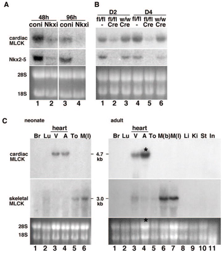
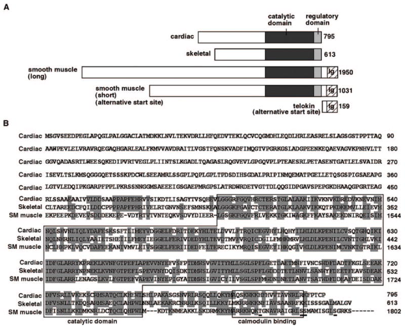
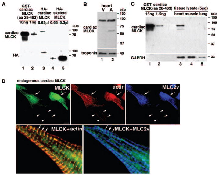
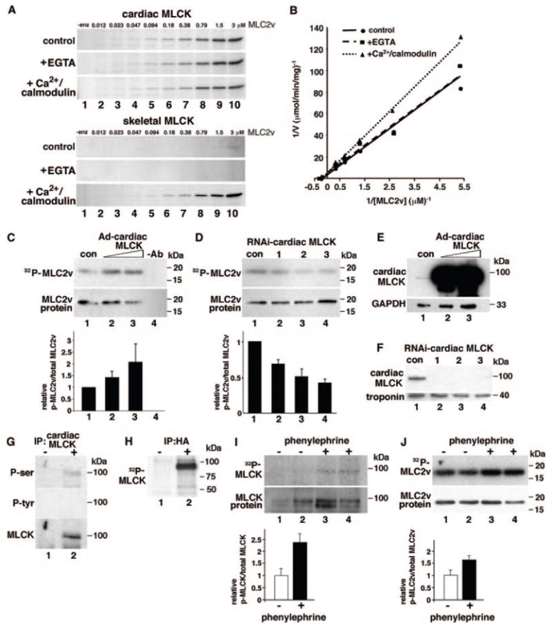
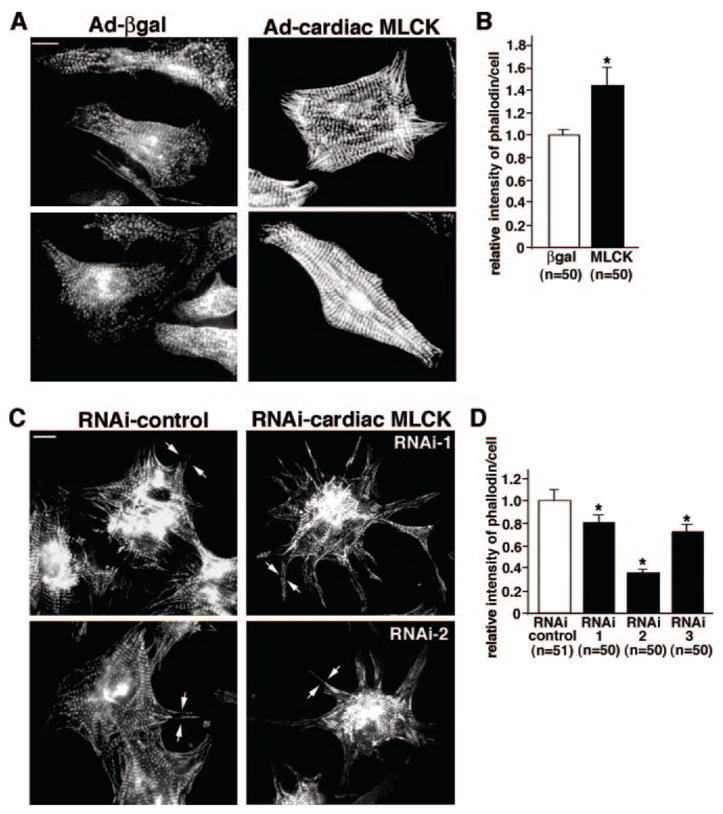
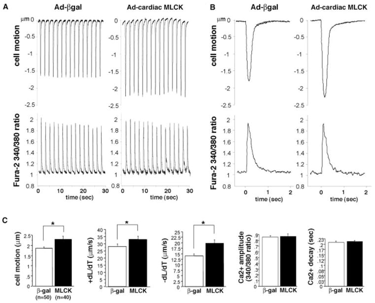
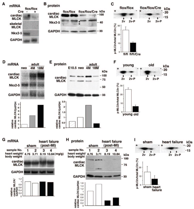
Two myosin light chain (MLC) kinase (MLCK) proteins, smooth muscle (encoded by mylk1 gene) and skeletal (encoded by mylk2 gene) MLCK, have been shown to be expressed in mammals. Even though phosphorylation of its putative substrate, MLC2, is recognized as a key regulator of cardiac contraction, a MLCK that is preferentially expressed in cardiac muscle has not yet been identified. In this study, we characterized a new kinase encoded by a gene homologous to mylk1 and -2, named cardiac MLCK, which is specifically expressed in the heart in both atrium and ventricle. In fact, expression of cardiac MLCK is highly regulated by the cardiac homeobox protein Nkx2-5 in neonatal cardiomyocytes. The overall structure of cardiac MLCK protein is conserved with skeletal and smooth muscle MLCK; however, the amino terminus is quite unique, without significant homology to other known proteins, and its catalytic activity does not appear to be regulated by Ca(2+)/calmodulin in vitro. Cardiac MLCK is phosphorylated and the level of phosphorylation is increased by phenylephrine stimulation accompanied by increased level of MLC2v phosphorylation. Both overexpression and knockdown of cardiac MLCK in cultured cardiomyocytes revealed that cardiac MLCK is likely a new regulator of MLC2 phosphorylation, sarcomere organization, and cardiomyocyte contraction.


Figures

Figure 1
Figure 1
Identification of cardiac-specific MLCK as an Nkx2-5 downstream target gene. A, Northern blotting showed that Nkx2-5 knockdown using adenovirus shRNA-reduced expression of cardiac MLCK 48 hours (lane 1 vs 2) and 96 hours after adenovirus infection (lane 3 vs 4). B, Tamoxifen-induced Nkx2-5 knockout demonstrates reduction of cardiac MLCK expression at postnatal day 2 (D2) (lane 2 vs lanes 1 and 3) and day 4 (D4) (lane 5 vs lanes 4 and 6). Of note, tamoxifen was injected into the pregnant female within 24 hours before delivery. Coni indicates control RNAi; Nkxi, Nkx2-5-RNAi; fl, floxed-Nkx2-5; fl/fl, homozygous for floxed-Nkx2-5; w, wild-type; w/w, homozygous for wild-type; and Cre, Cre-transgene. C, Tissue-specific expression of cardiac MLCK mRNA was examined by Northern blotting (top gels) and compared with that of skeletal MLCK (middle gels) in the neonatal stage (left) and adult stage (right). Increased loading of RNA isolated from adult atrium resulted in higher expression of MLCK in adult atrium vs ventricle (*, lane 4, right). Br indicates brain; Lu, lung; V, ventricle; A, atrium; To, tong; M(l), leg muscle; M(b), back muscle; Li, liver; Ki, kidney; St, stomach; and In, intestine.
Figure 2
Figure 2
Structure of mouse cardiac MLCK (GenBank accession number EU403565) protein compared with skeletal and smooth muscle MLCKs. A, Schematic of cardiac MLCK protein structure compared with skeletal and long and short forms of smooth muscle MLCK and variant of smooth muscle MLCK, telokin. Protein sequences are retrieved from mouse skeletal MLCK (XP_979674), smooth muscle MLCK (long) (NP647461), smooth muscle MLCK (short), and telokin (AAG34169). Ig indicates immunoglobulin C2 like motif. B, Amino acid sequence of cardiac MLCK with alignment among cardiac, skeletal, and smooth muscle (long-form) MLCK at the carboxyl terminus. Identical amino acids between at 2 proteins are shaded. Putative Ca2+/calmodulin binding kinase regulatory domain locating carboxyl terminus to catalytic domain; 2 contiguous serine residues, which are targets of upstream kinases and 2 additional autophosphorylation sites identified in the smooth muscle MLCK are underlined.
Figure 3
Figure 3
Cardiac MLCK protein expression and intracellular localization. A, Western blotting using anti–cardiac MLCK antibody against GST–cardiac MLCK (amino acids 28 to 463) (lanes 1 and 2) detected HA-tagged full-length cardiac MLCK protein (lane 3) but not HA-tagged full-length skeletal MLCK (lanes 4 and 5). Anti-HA antibody recognizes HA-tagged cardiac and skeletal MLCK (lanes 3 to 5). B, Western blotting using anti–cardiac MLCK antibody detected a similar amount of MLCK protein in protein lysates isolated from mouse neonatal ventricles (lane 1) and atria (lane 2). V indicates ventricle; A, atrium. C, Cardiac MLCK protein expression in heart lysates is equivalent to 2.3 ng of GST–cardiac MLCK in 5 μg of heart lysates. The antibody does not cross-react with other proteins using similar amounts of tissue lysates from skeletal muscle and lung (lanes 4 and 5). D, Coimmunostaining of cardiac MLCK, actin (phalloidin), and MLC2v. Endogenous cardiac MLCK protein is localized diffusely in the cytoplasm with occasional striated patterns (arrows) that overlap with actin but not with striated MLC2v. Arrowheads indicate noncardiomyocyte weakly detected by phalloidin but not by cardiac MLCK nor MLC2 antibodies. Bars=10 μm.
Figure 4
Figure 4
Cardiac MLCK phosphorylates MLC2 and potentially MLCK itself. A, Autoradiogram of GST-MLC2v phosphorylation at various concentrations (0.012 to 3 μmol/L) catalyzed by cardiac MLCK (upper gels) and skeletal MLCK (lower gels). Control experiments without EGTA or Ca2+/calmodulin (control), with EGTA (+EGTA, 5 mmol/L), or with Ca2+/calmodulin (+Ca2+/calmodulin, 1 mmol/L Ca2+, and 1 μmol/L calmodulin) are shown. B, Kinetic analysis of cardiac MLCK using double reciprocal Lineweaver–Burk plot (0.18 to 3 μmol/L). Calculated kinetic values of cardiac MLCK were as follows: Km (μmol/L) 4.3±1.5 (without EGTA or Ca2+/calmodulin), 2.9±0.8 (with EGTA), and 3.9±1.2 (with Ca2+/calmodulin); Vmax (μmol/min per milligram): 0.26±0.06, (without EGTA or Ca2+/calmodulin), 0.18±0.03 (with EGTA), and 0.18±0.03 (with Ca2+/calmodulin). Values are expressed as means±SE (n=2). C, MLC2v phosphorylation was examined in neonatal cardiomyocytes with increased expression of cardiac MLCK by infection of Ad-cardiac MLCK (lanes 2, 3 vs lane 1). The relative amounts of phosphorylated to total MLC2v protein are shown below in comparison with control cardiomyocytes. Lane 1: Ad-βgal, 10 multiplicities of infection (mois); lane 2: Ad-cardiac MLCK, 2 mois; lane 3: Ad-cardiac MLCK, 10 mois. The relative values are expressed as means±SE (n=2). D, Decreased expression of cardiac MLCK using 3 different shRNAs (lanes 2 to 4) decreased MLC2v phosphorylation compared with scrambled adenovirus-shRNA (lane 1). The relative values of phosphorylated to total MLC2v protein in comparison with control cardiomyocytes are expressed as means±SE (n=4). E, Increased expression of cardiac MLCK by infection of Ad-cardiac MLCK adenovirus detected by Western blotting with anti–cardiac MLCK antibody. Lane 1: Ad-βgal, 10 mois; lane 2: Ad-cardiac MLCK, 2 mois; lane 3: Ad-cardiac MLCK, 10 mois. F, Reduced expression of cardiac MLCK by 3 different shRNA is detected by Western blotting with anti–cardiac MLCK antibody. Lane 1, shRNA with scrambled sequence; lanes 2 to 4, shRNA targeting to 3 different sequences. G, Cardiac MLCK immunoprecipitated from neonatal ventricular cardiomyocytes was blotted with anti–phospho-serine or –phospho-tyrosine antibodies. Phospho-serine antibody reacted to cardiac MLCK protein. H, In vitro kinase assay showed 32P-incorporated HA-tagged MLCK expressed in 293 cells. I, Phenylephrine (30 μmol/L for 30 minutes) increased cardiac MLCK phosphorylation in cardiomyocytes infected with Ad-HA cardiac MLCK (1 moi). The relative amounts of phosphorylated cardiac MLCK to total cardiac MLCK protein are shown below in comparison with control cardiomyocytes without phenylephrine treatments (mean±SE, n=2). J, Phenylephrine (30 μmol/L for 30 minutes) increased cardiac MLC2v phosphorylation in rat neonatal cardiomyocytes. The relative values of phosphorylated to total MLC2v protein in comparison with control cardiomyocytes without phenylephrine are shown (mean±SE, n=2).
Figure 5
Figure 5
Effects of cardiac MLCK in sarcomere organization. A, Overexpression of cardiac MLCK by Ad-cardiac MLCK adenovirus (10 mois) promotes sarcomere organization detected by phalloidin compared with cells infected with Ad-βgal (10 mois). Bars=10 μm. B, Relative intensity of phalloidin staining in individual cardiomyocytes overexpressing cardiac MLCK is shown with the value in control cardiomyocytes defined as 1 (mean±SE). C, Reduced expression of cardiac MLCK using 2 different shRNAs, RNAi-1 and RNAi-2, does not disturb sarcomere organization centrally with slight changes in peripheral structure (arrows). Bars=10 μm. D, Relative intensity of phalloidin staining in individual cardiomyocyte treated with 3 different shRNAs is shown with the value in control cardiomyocytes defined as 1 (mean±SE).
Figure 6
Figure 6
Cardiac MLCK potentiates cardiomyocyte contraction. A, Representative tracings of cell motion (top) and simultaneous Ca2+ transients (bottom) in an isolated rat neonatal cardiomyocytes obtained continuously for 30 seconds. Ten multiplicities of infection of adenovirus encoding βgal or cardiac MLCK were infected. B, Representative tracings of a single contraction are shown. C, Summarized data obtained from multiple cardiomyocytes demonstrate that increased cardiac MLCK expression significantly increases cell motion, +dL/dT (contraction) and −dL/dT (relaxation), without a significant change in Ca2+ amplitude and decay. Values are expressed as means±SE.
Figure 7
Figure 7
Cardiac MLCK expression and MLC2v phosphorylation in mice with Nkx2-5 knockout, aging, and post–myocardial infarction (MI) heart failure. A and B, Decreased expression of cardiac MLCK mRNA and protein in Nkx2-5 knockout hearts at postnatal day 12. Skeletal MLCK mRNA was not detected by Northern blotting. C, Unphosphorylated (left, with higher pI) and phosphorylated (right, with lower pI) MLC2v examined in 2D electrophoresis, followed by Western blotting with anti-MLC antibody. Relative amounts of phosphorylated to total MLC2v are shown (mean±SE, n=2). D, Expression of cardiac MLCK and Nkx2-5 mRNA in neonatal, adult (4 months) and aged hearts (18 months). Relative expression of cardiac MLCK normalized to GAPDH is shown with the value in neonatal heart defined as 1. E, Expression of cardiac MLCK protein in embryonic day 10.5, neonatal, adult, and aged hearts. Relative expression of cardiac MLCK normalized to GAPDH is shown with the value in neonatal heart defined as 1. F, Level of MLC2v phosphorylation in young (PD 12) and aged hearts. Relative amounts of phosphorylated to total MLC2v are shown (mean±SE; young, n=3; old, n=6 from 2 mice at 18 and 21 months). G, Noninfarcted upper septal tissue dissected from mice 3 weeks after coronary ligation (3 month old) was analyzed for cardiac MLCK mRNA expression: 2 sham-operated (lanes 1, 2) and 2 heart failure (lanes 3 and 4) mice. Values of heart weight/body weight are indicated. Additional experimental conditions and parameters of cardiac function have been described previously and in Materials and Methods. Relative expression of cardiac MLCK normalized to GAPDH is shown with the value in sample 1 defined as 1. H, Cardiac MLCK protein expression in tissue lysates from the same mice used in G is shown with the value in sample 1 defined as 1. I, Level of MLC2v phosphorylation examined in mice with sham-operated and heart failure. Relative amounts of phosphorylated to total MLC2v are shown (mean±SE; sham, n=4 from 2 mice; heart failure, n=4 from 2 mice).

Comment in

Similar articles

Cited by

References

    1. Alberts B, Johnson A, Lewis J, Raff M, Roberts K, Walter P. Molecular Biology of the Cell. 4. New York: Garland Publishing Inc; 2002.
    1. Murthy KS. Signaling for contraction and relaxation in smooth muscle of the gut. Annu Rev Physiol. 2006;68:345–374. - PubMed
    1. Kamm KE, Stull JT. Dedicated myosin light chain kinases with diverse cellular functions. J Biol Chem. 2001;276:4527–4530. - PubMed
    1. Stelzer JE, Patel JR, Moss RL. Acceleration of stretch activation in murine myocardium due to phosphorylation of myosin regulatory light chain. J Gen Physiol. 2006;128:261–272. - PMC - PubMed
    1. Moss RL, Fitzsimons DP. Myosin light chain 2 into the mainstream of cardiac development and contractility. Circ Res. 2006;99:225–227. - PubMed

Publication types

MeSH terms

Associated data