. 2008 Jan;178(1):513-26.
doi: 10.1534/genetics.107.083253.

Distinct activities of the germline and somatic reproductive tissues in the regulation of Caenorhabditis elegans' longevity

Affiliations

Distinct activities of the germline and somatic reproductive tissues in the regulation of Caenorhabditis elegans' longevity

Tracy M Yamawaki et al. Genetics. 2008 Jan.

Abstract

The two parts of the Caenorhabditis elegans reproductive system, the germ cells and the somatic reproductive tissues, each influence the life span of the animal. Removing the germ cells increases longevity, and this life span extension requires the somatic gonad. Here we show that the somatic gonad and the germ cells make distinct contributions to life span determination. The life span increase produced by loss of the germ cells requires the DAF-16/FOXO transcription factor. In response to germ-cell removal, DAF-16 accumulates in nuclei. We find that the somatic gonad is not required for DAF-16 nuclear accumulation or for the increased stress resistance that is produced by germ-cell removal. The somatic gonad is required, however, for expression of specific DAF-16 target genes. DAF-16 is known to be activated by reduced insulin/IGF-1 signaling in C. elegans. In certain insulin/IGF-1-pathway mutants, the somatic gonad is not required for germ-cell removal to extend life span. Our genetic experiments suggest that these mutations reduce insulin/IGF-1 signaling below a critical threshold level. At these low levels of insulin/IGF-1 signaling, factors normally provided by the somatic gonad are no longer needed for germ-cell removal to increase the expression of DAF-16 target genes.


Figures
F<sc>igure</sc> 1.—
Figure 1.—
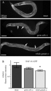
The effects of somatic-gonad removal on the pattern of DAF-16∷GFP. (A) The somatic gonad is not required for DAF-16∷GFP nuclear localization in animals that lack germ cells. Arrows indicate nuclear localization of DAF-16∷GFP in intestinal cells of day-2 adults lacking the germ cells (Z2 and Z3 ablated at hatching) or the germ cells as well as the somatic gonad (Z1 and Z4 ablated at hatching). Approximately 100 animals were examined in multiple trials, and the animals shown are representative. Nuclear localization of DAF-16∷GFP was observed in all of the animals lacking either germ cells or the whole gonad. (B) Somatic-gonad removal affects the level of DAF-16∷GFP. Removing the somatic gonad produced a modest but statistically significant decrease in the level of DAF-16∷GFP fluorescence. CF1934 intact control, n = 8, m = 1 ± 0.11; Z2/3, n = 14, m = 0.87 ± 0.037, P = 0.28; Z1/4, n = 6, m = 0.76 ± 0.028, P = 0.072, P′ = 0.039. Mean fluorescence intensity given is relative to intact control. P, the P-value (Student's t-test) compared to intact. P′, the P-value comparing germ-cell (Z2/Z3) ablation to whole-gonad (Z1/Z4) ablation.
F<sc>igure</sc> 2.—
Figure 2.—
The somatic gonad is required for expression of some DAF-16-regulated genes in animals that lack germ cells. (A) Expression of GFP reporters in animals lacking germ cells (Z2 and Z3 ablated) or the somatic gonad and germ cells (Z1 and Z4 ablated). Values for histograms are given in Table 1. Psod-3∷gfp and Pgpd-2∷gfp expression requires the presence of the somatic gonad in germ-cell ablated animals. In five of nine trials, there was a statistically significant decrease in expression of dod-8 in whole-gonad ablated animals. Trial 1, shown here, is representative of this observation. In four of nine trials (such as trial 4, displayed here) there was no significant decrease in dod-8 expression upon whole gonad ablation. In one of four trials (trial 3, shown here) there was a statistically significant decrease in expression of nnt-1 when the somatic gonad as well as the germ cells were removed (Z1 and Z4 ablated). In three of four trials (such as trial 1, shown here) there was no significant decrease in nnt-1 expression upon whole gonad ablation. (B) Expression of GFP reporters in daf-16(mu86) mutants lacking either the germ cells (Z2 and Z3 ablated) or the germ cells and the somatic gonad (Z1 and Z4 ablated). Thus, the increase in Psod-3∷gfp and Pgpd-2∷gfp expression produced by germline removal requires daf-16 and the somatic gonad. In contrast, the increase in Pdod-8∷gfp and Pnnt-1∷gfp expression is partially daf-16 and somatic-gonad independent. For complete data set, see Table 2.
F<sc>igure</sc> 3.—
Figure 3.—
The reproductive system and insulin-like signaling. (A) The somatic gonad is required for the life span extension produced by germline removal in weak insulin/IGF-1-pathway mutants. daf-2(RNAi) intact control, n = 128/113 (uncensored/total analyzed), m = 36.5 ± 1.7 (days); Z2/3(−), n = 90/73, m = 72.8 ± 2.5, P < 0.001; Z1/4(−), n = 93/80, m = 58.2 ± 2.2, P < 0.0001, P′ < 0.0001. pdk-1(sa709) intact control, n = 64/34, m = 26.6 ± 1.2; Z2/3(−), n = 44/37, m = 33.7 ± 2.0, P = 0.0026; Z1/4(−), n = 48/45, m = 28.0 ± 0.98, P = 0.54, P′ = 0.0015. akt-1(ok525) intact control, n = 90/71, m = 21.3 ± 0.73; Z2/3(−), n = 94/46, m = 28.0 ± 1.6, P < 0.0001; Z1/4(−), n = 88/85, m = 23.9 ± 1.1, P = 0.015, P′ = 0.048; akt-1(+), n = 90/72, m = 16.7 ± 0.53, P < 0.0001. akt-2(ok393) intact control, n = 90/64, m = 14.0 ± 0.61; Z2/3(−), n = 88/57, m = 19.8 ± 1.3, P < 0.0001; Z1/4(−), n = 104/96, m = 17.3 ± 0.54, P = 0.0001, P′ = 0.0363; akt-2(+), n = 81/61, m = 14.9 ± 0.55, P = 0.48. P refers to the P-value compared to intact. P′ refers to the P-value comparing germ-cell (Z2/Z3) ablation to whole-gonad (Z1/Z4) ablation. (B) Reducing daf-2 levels in daf-2(mu150) mutants with RNAi allows germline removal to extend life span independently of the somatic gonad. daf-2(mu150) fed HT115 bacteria carrying the pAD12 vector only control plasmid: intact control, n = 79/61, m = 27.0 ± 0.862; Z2/3(−), n = 48/34, m = 44.0 ± 2.7, P <0.001; Z1/4(−), n = 52/51, m = 28.8 ± 1.0, P = 0.12, P′ < 0.0001. daf-2(mu150, RNAi) fed HT115 bacteria carrying the pAD43 (daf-2 RNAi) plasmid: intact control, n = 77/61, m = 49.1 ± 1.9; Z2/3(−), n = 46/39, m = 92.3 ± 4.5, P < 0.0001; Z1/4(−), n = 51/45, m = 102.4 ± 3.4, P < 0.0001, P′ = 0.27. (C) Quantitative model to explain the difference in life span seen with whole gonad ablation in various daf-2 mutant backgrounds. Decreasing the amount of insulin-like signaling below a certain threshold eliminates the requirement for the somatic gonad.
F<sc>igure</sc> 4.—
Figure 4.—
Expression of the DAF-16 target gene sod-3 generally correlates with life span in daf-2 mutants lacking either the germ cells or the somatic gonad. Fluorescence intensity of GFP from the whole animal, excluding vulval regions, was measured and calculated relative to intact controls (see materials and methods). Values for histograms of sod-3∷gfp levels are given in Table 4.
F<sc>igure</sc> 5.—
Figure 5.—
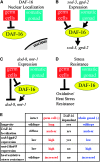
The somatic reproductive tissues are required for some, but not all, of the processes triggered by germline removal. (A) Loss of the germ cells triggers DAF-16 nuclear localization independently of the somatic gonad. (B) The somatic gonad is required for the increased expression of sod-3 and gpd-2 that occurs when the germ cells are removed. (C) The somatic gonad is not required for the increased expression of dod-8 and nnt-1 that occurs when the germ cells are removed. Moreover, DAF-16 is only partially required for this upregulation. (D) Neither DAF-16 nor the somatic gonad is required for the increased stress resistance that occurs when the germ cells are removed. (E) Specific findings demonstrating that the somatic gonad is required for some, but not all processes triggered by germ-cell removal.

Similar articles

Cited by

References

    1. Arantes-Oliveira, N., J. Apfeld, A. Dillin and C. Kenyon, 2002. Regulation of life-span by germ-line stem cells in Caenorhabditis elegans. Science 295: 502–505. - PubMed
    1. Arantes-Oliveira, N., J. R. Berman and C. Kenyon, 2003. Healthy animals with extreme longevity. Science 302: 611. - PubMed
    1. Berman, J. R., and C. Kenyon, 2006. Germ-cell loss extends C. elegans life span through regulation of DAF-16 by kri-1 and lipophilic-hormone signaling. Cell 124: 1055–1068. - PubMed
    1. Brenner, S., 1974. The genetics of Caenorhabditis elegans. Genetics 77: 71–94. - PMC - PubMed
    1. Broue, F., P. Liere, C. Kenyon and E. E. Baulieu, 2007. A steroid hormone that extends the lifespan of Caenorhabditis elegans. Aging Cell 6: 87–94. - PubMed

Publication types

MeSH terms