Review
. 2008 Jan;21(1):13-25.
doi: 10.1128/CMR.00022-07.

Sensing of viral infection and activation of innate immunity by toll-like receptor 3

Affiliations
Review

Sensing of viral infection and activation of innate immunity by toll-like receptor 3

Elisabeth Vercammen et al. Clin Microbiol Rev. 2008 Jan.

Abstract

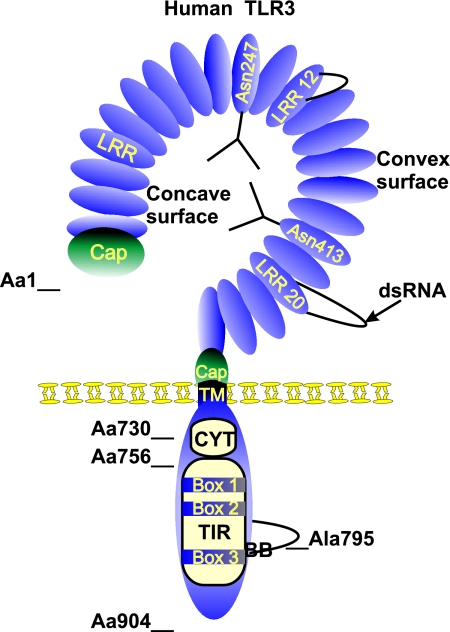
Toll-like receptors (TLRs) form a major group of transmembrane receptors that are involved in the detection of invading pathogens. Double-stranded RNA is a marker for viral infection that is recognized by TLR3. TLR3 triggering activates specific signaling pathways that culminate in the activation of NF-kappaB and IRF3 transcription factors, as well as apoptosis, enabling the host to mount an effective innate immune response through the induction of cytokines, chemokines, and other proinflammatory mediators. In this review, we describe the paradoxical role of TLR3 in innate immunity against different viruses and in viral pathogenesis but also the evidence for TLR3 as a "danger" receptor in nonviral diseases. We also discuss the structure and cellular localization of TLR3, as well as the complex signaling and regulatory events that contribute to TLR3-mediated immune responses.


Figures

FIG. 1.
FIG. 1.
Schematic structure of human TLR3. TLR3 is a type I integral membrane protein of 904 amino acids. The TLR3 extracellular domain is a horseshoe-shaped solenoid in which LRR forms one turn of the solenoid. The LRRs are at the N-terminal and C-terminal regions, flanked by a cysteine-rich Cap domain. The concave surface is rich in potential N-glycosylation sites and probably heavily glycosylated. Here we represent two N-glycan structures on Asn247 and Asn413, two residues which are implicated in glycosylation. LRR12 and LRR20 are atypical LRR motifs containing large insertions which protrude from the solenoid. According to the symmetrical assembly model, ligand binding occurs at the glycan-free surface involving LRR20. The transmembrane domain (TM) is made up of one single α-helix. The cytoplasmic domain comprises the cytoplasmic linker region (CYT) (amino acid [Aa] 730 to Aa756) and the TIR domain, from which the adaptor-binding BB loop protrudes. Ala795 is a conserved residue residing at the top of the BB loop and is involved in the binding of TRIF. The three conserved boxes that define the TIR domain are also indicated.
FIG. 2.
FIG. 2.
TLR3 signaling pathways. Binding of dsRNA to the TLR3-CD14 complex induces the activation of several intracellular signaling pathways. The activation of NF-κB and IRF3 is achieved by two different signaling branches emanating from the TLR3 adaptor molecule TRIF, which binds to the BB loop of the TLR3 TIR domain. Distinct regions of TRIF bind the ubiquitin ligase TRAF6 and the kinase RIP1. Analogously with the ubiquitin ligase activity of TRAF2 in the TNF receptor pathway, the activity of TRIF-associated TRAF6 might be responsible for the Ub of RIP1 in the TLR3 pathway. RIP1 ubiquitination is recognized by the ubiquitin receptor proteins TAB2 and TAB3, leading to the activation of the kinase TAK1, which is part of the same complex. TAK1 phosphorylates and activates IKKα and IKKβ, which are part of a bigger IKK complex with the IKK adaptor protein IKKγ. IKKβ is known to be the crucial IKK in TLR signaling and phosphorylates IκBα, which binds and keeps NF-κB (here depicted as a p65/p50 dimer) in an inactive state in the cytoplasm. IκBα phosphorylation leads to its recognition and degradation by the proteasome, thus allowing NF-κB to translocate to the nucleus, where it binds and activates specific gene promoters (e.g., A20). TRIF also binds TRAF3 and NAP1. Whereas the role of TRAF3 is still largely unclear, NAP1 functions as an adaptor for the IKK-related kinases IKKɛ and TBK1, which have largely redundant functions. Both kinases phosphorylate IRF3, leading to its dimerization and translocation to the nucleus, where it binds and activates specific gene promoters (e.g., IFN-β). Whereas these TRIF-mediated signaling pathways result in the activation of NF-κB and IRF3, the phosphorylation of NF-κB and IRF3 is involved in acquiring the fully activated status of both transcription factors (see the text for more details). Signaling leading to these events is still largely unclear, but IRF3 phosphorylation is dependent on the kinase Akt, which is activated by the lipid kinase PI3K, which binds phospho-Tyr759 of TLR3. Interestingly, PI3K also seems to have an inhibitory function on NF-κB activation, whereas the phosphorylation of TLR3 on Tyr858 enhances NF-κB activation by an unknown mechanism. TLR3 also induces apoptosis via a TRIF- and RIP1-dependent mechanism. The binding of RIP1 to TRIF not only activates NF-κB but also recruits the DD-containing adaptor protein FADD via a homotypic DD-DD interaction. FADD in turn interacts with the cysteine protease procaspase-8 through the death effector domain (DED) present in both proteins. This is believed to result in the proteolytic auto-activation of procaspase-8 and the initiation of cell death. CYT, cytoplasmic linker.
FIG. 3.
FIG. 3.
Endogenous and viral (green) inhibitors of TLR3-mediated NF-κB or IRF3 activation. Most known inhibitors interfere with the function of TRIF, either by interacting with TRIF (PIASy, TRAF1, SARM, A20, TRAF4, and the vaccinia virus protein A46R) or by degrading TRIF (hepatitis C virus protease NS3/4A). Other inhibitors interact with TRAF6 (TRAF4, A20, and vaccinia virus protein A52R), RIP1 (RIP3), TBK1/IKKɛ (A20, SIKE, and SHP-2), or IRF3 (PIASy). See the text for more details.

References

    1. Abujamra, A. L., R. A. Spanjaard, I. Akinsheye, X. Zhao, D. V. Faller, and S. K. Ghosh. 2006. Leukemia virus long terminal repeat activates NFκB pathway by a TLR3-dependent mechanism. Virology 345:390-403. - PMC - PubMed
    1. Adis, R. D. P. 2004. Mismatched double-stranded RNA: polyI:polyC12U. Drugs R D 5:297-304. - PMC - PubMed
    1. Aksoy, E., W. Vanden Berghe, S. Detienne, Z. Amraoui, K. A. Fitzgerald, G. Haegeman, M. Goldman, and F. Willems. 2005. Inhibition of phosphoinositide 3-kinase enhances TRIF-dependent NF-κB activation and IFN-β synthesis downstream of Toll-like receptor 3 and 4. Eur. J. Immunol. 35:2200-2209. - PubMed
    1. Alexopoulou, L., A. C. Holt, R. Medzhitov, and R. A. Flavell. 2001. Recognition of double-stranded RNA and activation of NF-κB by Toll-like receptor 3. Nature 413:732-738. - PubMed
    1. An, H., W. Zhao, J. Hou, Y. Zhang, Y. Xie, Y. Zheng, H. Xu, C. Qian, J. Zhou, Y. Yu, S. Liu, G. Feng, and X. Cao. 2006. SHP-2 phosphatase negatively regulates the TRIF adaptor protein-dependent type I interferon and proinflammatory cytokine production. Immunity 25:919-928. - PubMed

Publication types

LinkOut - more resources