Review
. 2008 Jan;21(1):26-59.
doi: 10.1128/CMR.00019-07.

Complicated catheter-associated urinary tract infections due to Escherichia coli and Proteus mirabilis

Affiliations
Review

Complicated catheter-associated urinary tract infections due to Escherichia coli and Proteus mirabilis

S M Jacobsen et al. Clin Microbiol Rev. 2008 Jan.

Abstract

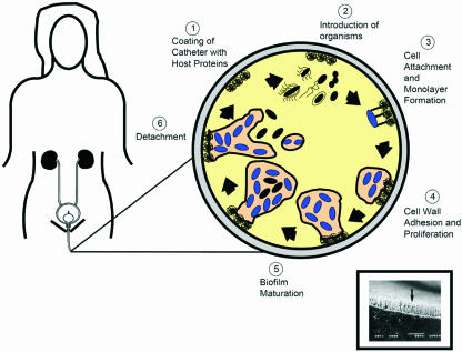
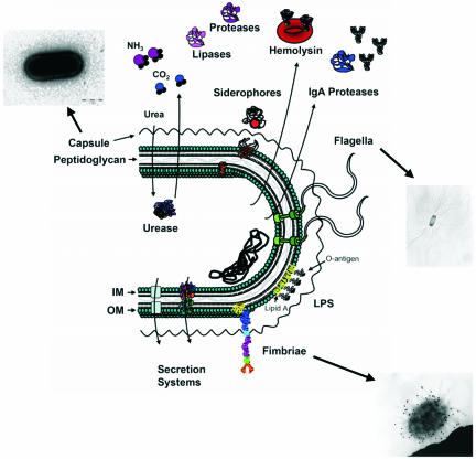
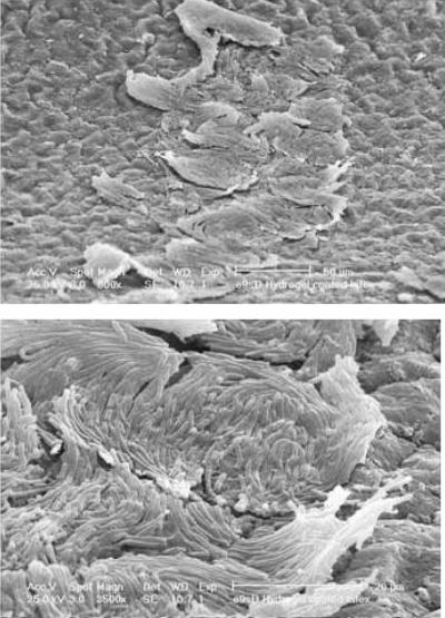
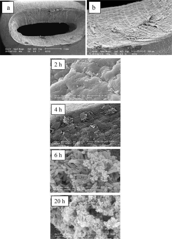
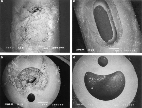
Catheter-associated urinary tract infections (CAUTIs) represent the most common type of nosocomial infection and are a major health concern due to the complications and frequent recurrence. These infections are often caused by Escherichia coli and Proteus mirabilis. Gram-negative bacterial species that cause CAUTIs express a number of virulence factors associated with adhesion, motility, biofilm formation, immunoavoidance, and nutrient acquisition as well as factors that cause damage to the host. These infections can be reduced by limiting catheter usage and ensuring that health care professionals correctly use closed-system Foley catheters. A number of novel approaches such as condom and suprapubic catheters, intermittent catheterization, new surfaces, catheters with antimicrobial agents, and probiotics have thus far met with limited success. While the diagnosis of symptomatic versus asymptomatic CAUTIs may be a contentious issue, it is generally agreed that once a catheterized patient is believed to have a symptomatic urinary tract infection, the catheter is removed if possible due to the high rate of relapse. Research focusing on the pathogenesis of CAUTIs will lead to a better understanding of the disease process and will subsequently lead to the development of new diagnosis, prevention, and treatment options.


Figures

FIG. 1.
FIG. 1.
Pathogenesis of biofilm formation on urinary catheters during CAUTIs. The inset (reprinted from reference 393) shows a scanning electron micrograph of a urinary catheter encrusted with P. aeruginosa.
FIG. 2.
FIG. 2.
Virulence factors of the gram-negative uropathogens E. coli and P. mirabilis. IM, inner membrane; OM, outer membrane. (The micrographs are reprinted from references , , and with permission.)
FIG. 3.
FIG. 3.
Cross section of a silicone catheter removed from a patient after blockage. Crystalline material can be seen completely occluding the catheter lumen. (Reprinted from reference 387a) with permission of the publisher.)
FIG. 4.
FIG. 4.
Crystalline material that blocked a patient's catheter after just 4 days. The large coffin-shaped crystals were shown by X-ray microanalysis to be a form of magnesium ammonium phosphate (struvite), and the microcrystalline aggregates were shown to be calcium phosphate (apatite). A four-membered bacterial community was isolated from this crystalline biofilm composed of E. coli, P. aeruginosa, E. faecalis, and P. mirabilis. (Modified from reference with permission from Elsevier.)
FIG. 5.
FIG. 5.
Scanning electron micrographs of a section of a hydrogel-coated latex catheter over which swarmer cells of P. mirabilis are migrating. The sections have been removed from the laboratory model described by Sabbuba et al. (337). Multicellular rafts of typical swarmer cells are visible on the irregular surface of the catheter, migrating from left to right. The micrographs were kindly provided by Rob Broomfield of Cardiff School of Biosciences, Cardiff University.
FIG. 6.
FIG. 6.
Early stages in the formation of a crystalline P. mirabilis biofilm on a hydrogel-coated latex catheter. Catheters were removed for examination by scanning electron microscopy after incubation for various times in a laboratory model of the catheterized bladder. The irregular nature of the surface of the eyelet is shown in a and b. After 2 h, in the model, cells can be seen trapped in crevices. At 4 h, microcolonies have formed in surface depressions. At 6 h, microcrystalline material accumulated in the developing biofilm as the pH of the urine rose. At 20 h, extensive crystalline biofilm formed at the eyehole. (Reproduced from reference with kind permission from Springer Science and Business Media.)
FIG. 7.
FIG. 7.
Scanning electron micrographs showing the extent of encrustation at the eyeholes and in the central channels of control (a and b) and triclosan-treated (c and d) silicone catheters. The control catheter was removed from the P. mirabilis-infected bladder model when it was blocked at 30 h. The triclosan-treated catheter drained freely for the experimental period and was removed from the model at 7 days. (Reprinted from reference with permission from Elsevier.)

References

    1. Abraham, S., J. Shin, and R. Malaviya. 2001. Type 1 fimbriated Escherichia coli-mast cell interactions in cystitis. J. Infect. Dis. 183(Suppl. 1):S51-S55. - PubMed
    1. Allison, C., L. Emody, N. Coleman, and C. Hughes. 1994. The role of swarm cell differentiation and multicellular migration in the uropathogenicity of Proteus mirabilis. J. Infect. Dis. 169:1155-1158. - PubMed
    1. Allison, C., H. C. Lai, D. Gygi, and C. Hughes. 1993. Cell differentiation of Proteus mirabilis is initiated by glutamine, a specific chemoattractant for swarming cells. Mol. Microbiol. 8:53-60. - PubMed
    1. Allison, C., H. C. Lai, and C. Hughes. 1992. Co-ordinate expression of virulence genes during swarm-cell differentiation and population migration of Proteus mirabilis. Mol. Microbiol. 6:1583-1591. - PubMed
    1. Altman, E., B. A. Harrison, R. K. Latta, K. K. Lee, J. F. Kelly, and P. Thibault. 2001. Galectin-3-mediated adherence of Proteus mirabilis to Madin-Darby canine kidney cells. Biochem. Cell Biol. 79:783-788. - PubMed

MeSH terms

Substances