

From clinical microbiology to infection pathogenesis: how daring to be different works for Staphylococcus lugdunensis
- PMID: 18202439
- PMCID: PMC2223846
- DOI: 10.1128/CMR.00036-07
From clinical microbiology to infection pathogenesis: how daring to be different works for Staphylococcus lugdunensis
Abstract
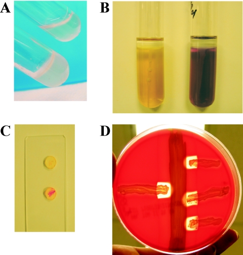
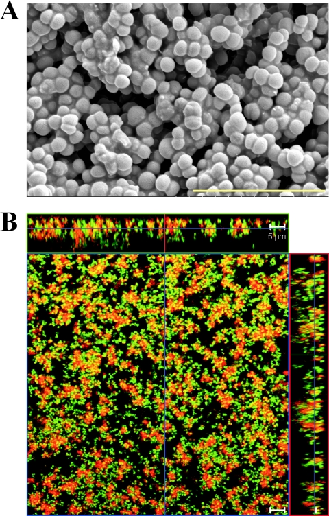
Staphylococcus lugdunensis has gained recognition as an atypically virulent pathogen with a unique microbiological and clinical profile. S. lugdunensis is coagulase negative due to the lack of production of secreted coagulase, but a membrane-bound form of the enzyme present in some isolates can result in misidentification of the organism as Staphylococcus aureus in the clinical microbiology laboratory. S. lugdunensis is a skin commensal and an infrequent pathogen compared to S. aureus and S. epidermidis, but clinically, infections caused by this organism resemble those caused by S. aureus rather than those caused by other coagulase-negative staphylococci. S. lugdunensis can cause acute and highly destructive cases of native valve endocarditis that often require surgical treatment in addition to antimicrobial therapy. Other types of S. lugdunensis infections include abscess and wound infection, urinary tract infection, and infection of intravascular catheters and other implanted medical devices. S. lugdunensis is generally susceptible to antimicrobial agents and shares CLSI antimicrobial susceptibility breakpoints with S. aureus. Virulence factors contributing to this organism's heightened pathogenicity remain largely unknown. Those characterized to date suggest that the organism has the ability to bind to and interact with host cells and to form biofilms on host tissues or prosthetic surfaces.
Figures
References
-
- Akiyama, H., H. Kanzaki, J. Tada, and J. Arata. 1998. Coagulase-negative staphylococci isolated from various skin lesions. J. Dermatol. 25:563-568. - PubMed
-
- Anguera, I., A. Del Rio, J. M. Miro, X. Matinez-Lacasa, F. Marco, J. R. Guma, G. Quaglio, X. Claramonte, A. Moreno, C. A. Mestres, E. Mauri, M. Azqueta, N. Benito, C. Garcia-de la Maria, M. Almela, M. J. Jimenez-Exposito, O. Sued, E. De Lazzari, and J. M. Gatell. 2005. Staphylococcus lugdunensis infective endocarditis: description of 10 cases and analysis of native valve, prosthetic valve, and pacemaker lead endocarditis clinical profiles. Heart 91:e10. - PMC - PubMed
-
- Arciola, C. R., D. Campoccia, Y. H. An, L. Baldassarri, V. Pirini, M. E. Donati, F. Pegreffi, and L. Montanaro. 2006. Prevalence and antibiotic resistance of 15 minor staphylococcal species colonizing orthopedic implants. Int. J. Artif. Organs. 29:395-401. - PubMed
-
- Asnis, D. S., S. St. John, R. Tickoo, and A. Arora. 2003. Staphylococcus lugdunensis breast abscess: is it real? Clin. Infect. Dis. 36:1348. - PubMed
-
- Bannerman, T. L., and S. J. Peacock. 2007. Staphylococcus, Micrococcus, and other catalase-positive cocci, p. 390-411. In P. R. Murray, E. J. Baron, J. H. Jorgensen, M. L. Landry, and M. A. Pfaller (ed.), Manual of clinical microbiology, 9th ed., vol. 1. ASM Press, Washington, DC.
Publication types
MeSH terms
Substances
LinkOut - more resources
Full Text Sources
Medical
Molecular Biology Databases
