Review
. 2008 Jan;21(1):134-56.
doi: 10.1128/CMR.00032-07.

Molecular pathogenesis of Shigella spp.: controlling host cell signaling, invasion, and death by type III secretion

Affiliations
Review

Molecular pathogenesis of Shigella spp.: controlling host cell signaling, invasion, and death by type III secretion

Gunnar N Schroeder et al. Clin Microbiol Rev. 2008 Jan.

Abstract

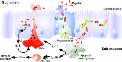
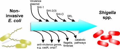
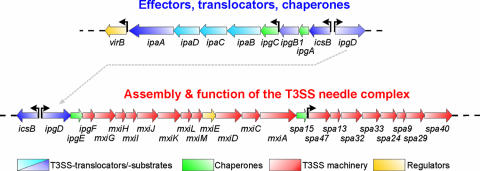
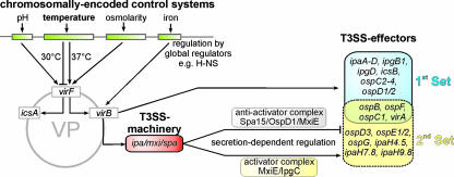
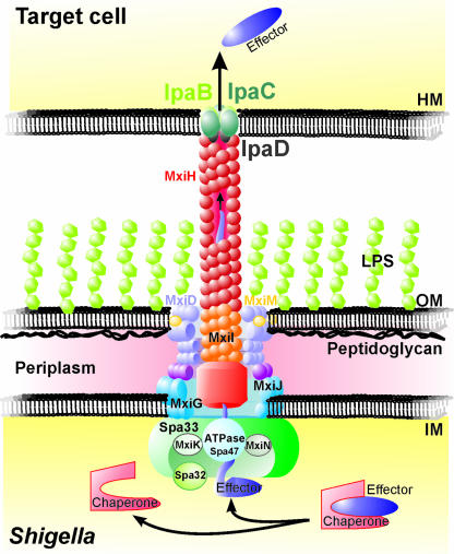
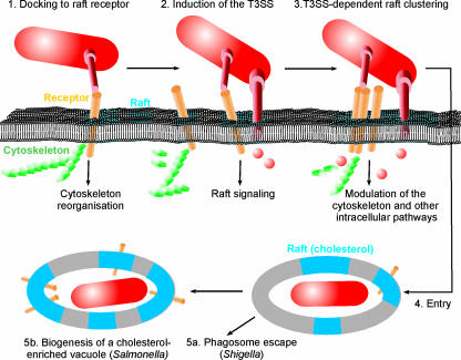
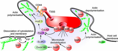
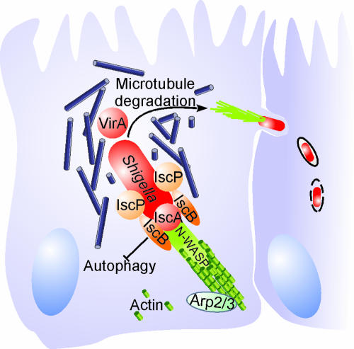
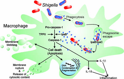
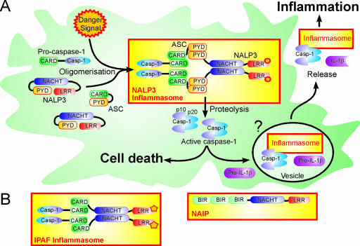
Shigella spp. are gram-negative pathogenic bacteria that evolved from harmless enterobacterial relatives and may cause devastating diarrhea upon ingestion. Research performed over the last 25 years revealed that a type III secretion system (T3SS) encoded on a large plasmid is a key virulence factor of Shigella flexneri. The T3SS determines the interactions of S. flexneri with intestinal cells by consecutively translocating two sets of effector proteins into the target cells. Thus, S. flexneri controls invasion into EC, intra- and intercellular spread, macrophage cell death, as well as host inflammatory responses. Some of the translocated effector proteins show novel biochemical activities by which they intercept host cell signal transduction pathways. An understanding of the molecular mechanisms underlying Shigella pathogenesis will foster the development of a safe and efficient vaccine, which, in parallel with improved hygiene, should curb infections by this widespread pathogen.


Figures

FIG. 1.
FIG. 1.
Cellular pathogenesis of Shigella spp. S. flexneri passes the EC barrier by transcytosis through M cells and encounters resident macrophages. The bacteria evade degradation in macrophages by inducing an apoptosis-like cell death, which is accompanied by proinflammatory signaling. Free bacteria invade the EC from the basolateral side, move into the cytoplasm by vectorial actin polymerization, and spread to adjacent cells. Proinflammatory signaling by macrophages and EC further activates the innate immune response involving NK cells and attracts PMN. The influx of PMN disintegrates the EC lining, which initially exacerbates the infection and tissue destruction by facilitating the invasion of more bacteria. Ultimately, PMN phagocytose and kill Shigella, thus contributing to the resolution of the infection.
FIG. 2.
FIG. 2.
Phylogenetic trees of Shigella and related enterobacteria. (A) Classical taxonomy of Shigella spp. based on phenotypic and numerical classification. (B) Evolutionary tree of Shigella and nonpathogenic E. coli strains (EcoR strains and K-12) based on comparative genomics. Shigella spp. fall into three main clusters, wherein the traditionally classified species are intermingled. The distribution of several E. coli EcoR strains between the Shigella strains indicates that the Shigella strains evolved from multiple ancestral origins. (Adapted from references and with permission from the Society for General Microbiology [United Kingdom] and Elsevier, respectively.)
FIG. 3.
FIG. 3.
Genetic events contributing to the evolution of Shigella spp. from nonpathogenic E. coli ancestors. Shigella spp. evolved from nonpathogenic E. coli through the acquisition of a large virulence plasmid and chromosomal pathogenicity islands as well as through the loss of genetic loci, which are not functional intracellularly or impede virulence. SRL, Shigella resistance locus. (Modified from reference with permission from Macmillan Publishers Ltd.)
FIG. 4.
FIG. 4.
Map of the 31-kb “entry region” on the S. flexneri virulence plasmid pWR100. The genes indicated encode structural components of the Mxi-Spa T3SS, secreted translocator and effector proteins, chaperones, and regulatory proteins. The region shown is essential and sufficient to invade EC and induce macrophage cell death.
FIG. 5.
FIG. 5.
Regulatory elements controlling the expression of the T3SS and its substrates on the S. flexneri virulence plasmid. The major virulence plasmid (VP) activator VirF is induced by environmental stimuli and triggers the expression of the central transcriptional activator VirB. VirB promotes the expression of the entry region genes and some additional effector genes scattered on the virulence plasmid. The secretion of the “first-set” T3SS effectors stored in the bacterial cytoplasm enables MxiE/IpgC-controlled induction of a discrete set of already-induced effector proteins and expression of the “second-set” effectors.
FIG. 6.
FIG. 6.
Architecture of the S. flexneri Mxi-Spa T3SS. The S. flexneri Mxi-Spa T3SS consists of four main parts. The seven-ringed basal body spans the bacterial IM, the periplasm, and the OM. The hollow needle is attached to a socket and protrudes from the basal body to the bacterial surface. Contact with host cell membranes (HM) triggers the IpaD-guided membrane insertion of the IpaB-IpaC translocon at the needle tip. The T3SS is completed by the cytoplasmic C ring, which is comprised of proteins that energize the transport process and mediate the recognition of substrates, chaperone release, and substrate unfolding.
FIG. 7.
FIG. 7.
Cholesterol and lipid rafts as triggers of type III secretion and targets of effectors. (1) The initial contact of bacteria with cholesterol-rich membrane microdomains is mediated by receptors, which partition to rafts. Receptor binding might already induce raft signaling, e.g., trigger the reorganization of the cytoskeleton. (2) Raft contact is followed by the cholesterol-dependent induction of effector protein secretion. (3) Receptor binding and effector secretion lead to the clustering of rafts and the subsequent uptake of the bacteria as well as the activation of other signaling pathways. (4) After uptake, the bacteria reside in a raft-containing vacuole, which is lysed (5a) or modified to create a replication-permissive niche (5b). Modification of the vacuole leads to a further enrichment of rafts (cholesterol), thus permitting bacterial manipulation of raft-associated signaling pathways.
FIG. 8.
FIG. 8.
Subversion of host cell signaling by S. flexneri type III-secreted effectors. Injection of the Mxi-Spa-secreted effectors IpaC, IpgB1, and VirA by S. flexneri induces Rac1/Cdc42-dependent actin polymerization and the formation of large membrane ruffles. The binding of IpaB to the CD44 receptor and the activity of IpgB2 might also trigger cytoskeleton remodeling or membrane ruffling, respectively. The phosphoinositide 4-phosphatase IpgD promotes the disconnection of the actin cytoskeleton from the cytoplasmic membrane, thus facilitating the structural reorganization of the entry site. IpaA mediates the localized depolymerization of actin, which is required to close the phagocytic cup. PIP2, phosphatidylinositol-4,5-biphosphate; PIP, phosphatidylinositol-5-phosphate.
FIG. 9.
FIG. 9.
Intracellular movement of S. flexneri by directed actin polymerization. Due to the activity of the serine protease SopA/IcsP, S. flexneri IcsA localizes to one pole of the bacterium, where it interacts with the host cell N-WASP protein. The IcsA/N-WASP complex recruits and activates the Arp2/Arp3 complex, thereby mediating actin nucleation. Elongation of the actin tail pushes S. flexneri through the cytoplasm. The movement is facilitated by VirA, which opens a path by degradation of the microtubule network. To avoid sequestration by the autophagy defense system, an autophagy recognition site on IcsA is masked by the protein IcsB.
FIG. 10.
FIG. 10.
S. flexneri-induced macrophage death. Depending on the Mxi-Spa T3SS, S. flexneri enhances its uptake by macrophages and secretes the translocators/effectors IpaB and IpaC to escape from the phagosome. In the cytoplasm, the remaining bacterial pool of IpaB is gradually released. Secreted IpaB integrates into membranes in a cholesterol-dependent manner and triggers the proteolytic activation of procaspase-1, which might be further promoted by TPPII. Active caspase-1 executes cell death and cleaves the precursors of the proinflammatory cytokines IL-1β and IL-18. The mature cytokines are released from the dying macrophage and elicit the strong inflammation characteristic of shigellosis.
FIG. 11.
FIG. 11.
Caspase-1 activation in the inflammasome. (A) Caspase-1 activation and IL-1β processing by the NALP3 inflammasome. A danger signal(s) induces a conformational change in NALP3, which leads to oligomerization and exposure of the PYD. Through homotypic PYD-PYD interactions, ASC is bound and recruits procaspase-1 through its CARD domain. Brought into close proximity, procaspase-1 undergoes autocatalytic cleavage into p10 and p20 fragments, which form the active caspase-1 tetramer. Active caspase-1 induces cell death and processes pro-IL-1β. The activation of caspase-1 occurs in or leads to the formation of vesicles through which caspase-1, IL-1β, and the inflammasome are released into the extracellular space. (B) Domain structure of the IPAF inflammasome and the NAIP.

References

    1. Abe, A., T. Matsuzawa, and A. Kuwae. 2005. Type-III effectors: sophisticated bacterial virulence factors. C. R. Biol. 328:413-428. - PubMed
    1. Adler, B., C. Sasakawa, T. Tobe, S. Makino, K. Komatsu, and M. Yoshikawa. 1989. A dual transcriptional activation system for the 230 kb plasmid genes coding for virulence-associated antigens of Shigella flexneri. Mol. Microbiol. 3:627-635. - PubMed
    1. Akeda, Y., and J. E. Galan. 2005. Chaperone release and unfolding of substrates in type III secretion. Nature 437:911-915. - PubMed
    1. Alder, N. N., and S. M. Theg. 2003. Energy use by biological protein transport pathways. Trends Biochem. Sci. 28:442-451. - PubMed
    1. Al-Hasani, K., I. R. Henderson, H. Sakellaris, K. Rajakumar, T. Grant, J. P. Nataro, R. Robins-Browne, and B. Adler. 2000. The sigA gene which is borne on the she pathogenicity island of Shigella flexneri 2a encodes an exported cytopathic protease involved in intestinal fluid accumulation. Infect. Immun. 68:2457-2463. - PMC - PubMed

Publication types

MeSH terms

Substances

LinkOut - more resources