Review
. 2008 Jan;21(1):157-97.
doi: 10.1128/CMR.00039-07.

Infections caused by Scedosporium spp

Affiliations
Review

Infections caused by Scedosporium spp

Karoll J Cortez et al. Clin Microbiol Rev. 2008 Jan.

Abstract

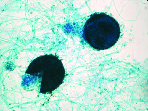
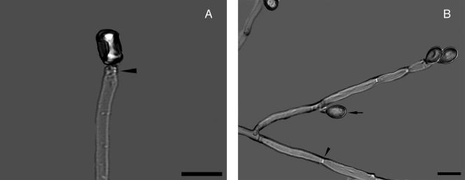
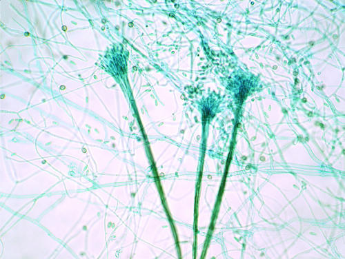
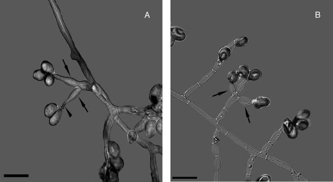
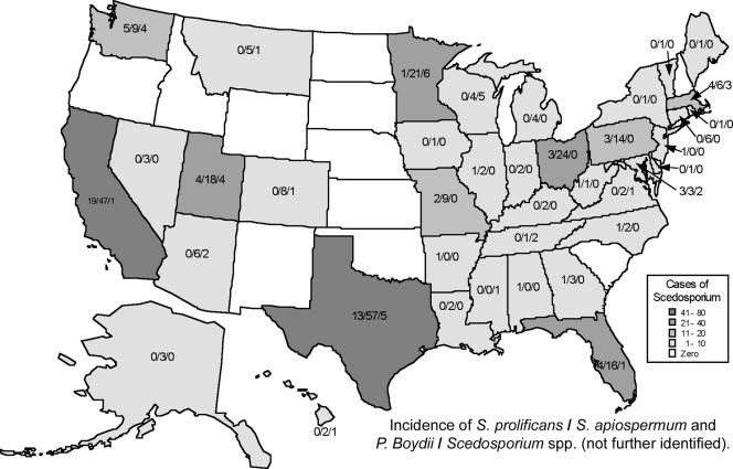
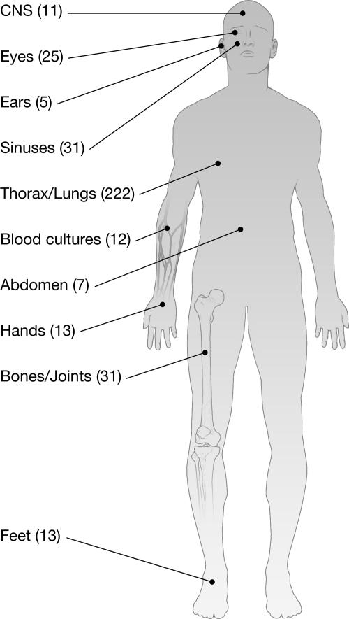
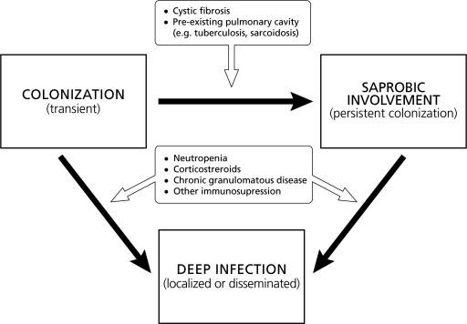
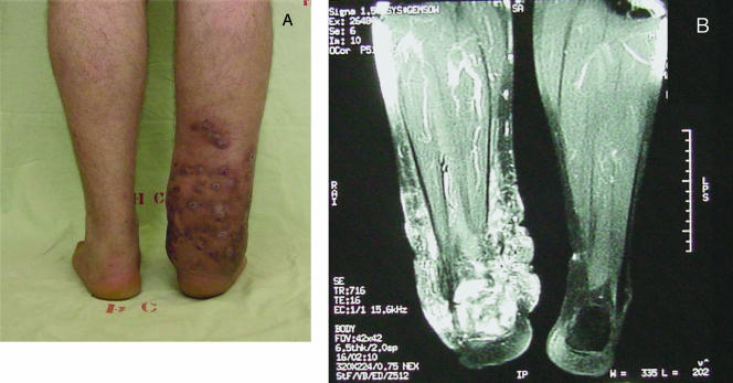
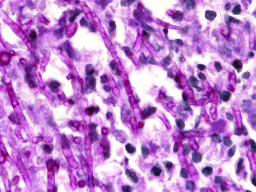
Scedosporium spp. are increasingly recognized as causes of resistant life-threatening infections in immunocompromised patients. Scedosporium spp. also cause a wide spectrum of conditions, including mycetoma, saprobic involvement and colonization of the airways, sinopulmonary infections, extrapulmonary localized infections, and disseminated infections. Invasive scedosporium infections are also associated with central nervous infection following near-drowning accidents. The most common sites of infection are the lungs, sinuses, bones, joints, eyes, and brain. Scedosporium apiospermum and Scedosporium prolificans are the two principal medically important species of this genus. Pseudallescheria boydii, the teleomorph of S. apiospermum, is recognized by the presence of cleistothecia. Recent advances in molecular taxonomy have advanced the understanding of the genus Scedosporium and have demonstrated a wider range of species than heretofore recognized. Studies of the pathogenesis of and immune response to Scedosporium spp. underscore the importance of innate host defenses in protection against these organisms. Microbiological diagnosis of Scedosporium spp. currently depends upon culture and morphological characterization. Molecular tools for clinical microbiological detection of Scedosporium spp. are currently investigational. Infections caused by S. apiospermum and P. boydii in patients and animals may respond to antifungal triazoles. By comparison, infections caused by S. prolificans seldom respond to medical therapy alone. Surgery and reversal of immunosuppression may be the only effective therapeutic options for infections caused by S. prolificans.


Figures

FIG. 1.
FIG. 1.
Pseudallescheria boydii in vitro, depicting a fully developed and ruptured cleistothecium, the hallmark of the sexual stage (teleomorph) of this fungus. Oblong ascospores are liberated in this culture. Magnification, ×100.
FIG. 2.
FIG. 2.
(A) Scedosporium apiospermum conidiophore with annellation (arrowhead). Note the solitary oval to pyriform conidium. (B) Acute-angle branching septate hyaline hyphae. Note the septum (arrowhead) and lateral conidiation (arrow). A KOH preparation using differential interference contrast with polarized light photographic technique is shown, Magnification, ×1,000; bar, 10 μm.
FIG. 3.
FIG. 3.
Synnemata (coremia) of the Graphium synanamorph of P. boydii bearing terminal conidia. Lactophenol cotton blue stain was used. Magnification, ×100.
FIG. 4.
FIG. 4.
Scedosporium prolificans (formerly Scedosporium inflatum). (A) The arrowhead points to annellations. The arrows point to the inflated shape of the conidiophores. (B) The arrows point to the inflated conidiophores generating pyriform conidia. A KOH preparation using differential interference contrast with polarized light photographic technique is shown. Bar, 10 μm.
FIG. 5.
FIG. 5.
Geographic distribution of cases which Scedosporium spp. were isolated in the United States from specimens submitted to the Fungus Testing Laboratory of the University of Texas Health Science System at San Antonio from January 2000 to May 2007. The white and gray tones represent the total incidences of Scedosporium cases reported by state. The numbers within each state indicate the incidence of Scedosporium prolificans/Scedosporium apiospermum and Pseudallescheria boydii/Scedosporium spp. (not further identified).
FIG. 6.
FIG. 6.
Anatomical origins (sites of infection) of 370 isolates submitted to the Fungus Testing Laboratory at the University of Texas Health Science System at San Antonio from January 2000 to May 2007.
FIG. 7.
FIG. 7.
Model of the host-pathogen interaction in pulmonary scedosporiosis. Pulmonary involvement begins with colonization of the respiratory tract. This colonization appears to be transient in immunocompetent hosts with anatomically normal respiratory tracts. However, colonization may become persistent in certain patient with anatomically altered respiratory tracts, leading to saprobic involvement. Such conditions occur in patients with cystic fibrosis, cavitary tuberculosis or sarcoidosis, and bronchiectasis. Conditions that alter the innate host defense mechanisms of a patient with colonization or saprobic involvement of the respiratory tract may lead to invasive disease manifesting as localized or disseminated infection. Conditions that may predispose to invasive pulmonary scedosporiosis include neutropenia, corticosteroid therapy, and CGD.
FIG. 8.
FIG. 8.
Scedosporium apiospermum pedal mycetoma of 18 years of evolution. Several sinus tracts in different evolution stages on the left foot (A) and draining white yellow grains resembling fig seeds at the openings of three fistulae (B) are shown. A transversal section of a fistula shows several lobed pale grains and an inflammatory infiltrate on the fistula lumen (C). H&E staining was used; magnification, ×400. (Reprinted from reference with permission from Elsevier.)
FIG. 9.
FIG. 9.
(A) Multifistulous right-lower-limb Scedosporium apiospermum mycetoma of 8 years of evolution. (B) MRI of the same patient, showing extensive inflammatory changes in the medial and lateral aspects of the lower leg and calcaneous osteomyelitis.
FIG. 10.
FIG. 10.
Scedosporium apiospermum infection in human tissue. The main host response is a mixed neutrophilic and monocytic infiltrate. PAS staining of brain tissue in a patient with CNS scedosporiosis was performed. Magnification, ×100.

References

    1. Abzug, M. J., and T. J. Walsh. 2004. Interferon-gamma and colony-stimulating factors as adjuvant therapy for refractory fungal infections in children. Pediatr. Infect. Dis. J. 23:769-773. - PubMed
    1. Acharya, A., A. Ghimire, B. Khanal, S. Bhattacharya, N. Kumari, and R. Kanungo. 2006. Brain abscess due to Scedosporium apiospermum in a non immunocompromised child. Indian J. Med. Microbiol. 24:231-232. - PubMed
    1. Adelson, H. T., and J. A. Malcolm. 1968. Endocavitary treatment of pulmonary mycetomas. Am. Rev. Respir. Dis. 98:87-92. - PubMed
    1. Afeltra, J., E. Dannaoui, J. F. Meis, J. L. Rodriguez-Tudela, and P. E. Verweij. 2002. In vitro synergistic interaction between amphotericin B and pentamidine against Scedosporium prolificans. Antimicrob. Agents Chemother. 46:3323-3326. - PMC - PubMed
    1. Ahmed, A. O., W. van Leeuwen, A. Fahal, W. van de Sande, H. Verbrugh, and A. van Belkum. 2004. Mycetoma caused by Madurella mycetomatis: a neglected infectious burden. Lancet Infect. Dis. 4:566-574. - PubMed

MeSH terms

LinkOut - more resources