. 2008 Jan 17;451(7176):345-9.
doi: 10.1038/nature06489.

Identification of cells initiating human melanomas

Affiliations

Identification of cells initiating human melanomas

Tobias Schatton et al. Nature. .

Abstract

Tumour-initiating cells capable of self-renewal and differentiation, which are responsible for tumour growth, have been identified in human haematological malignancies and solid cancers. If such minority populations are associated with tumour progression in human patients, specific targeting of tumour-initiating cells could be a strategy to eradicate cancers currently resistant to systemic therapy. Here we identify a subpopulation enriched for human malignant-melanoma-initiating cells (MMIC) defined by expression of the chemoresistance mediator ABCB5 (refs 7, 8) and show that specific targeting of this tumorigenic minority population inhibits tumour growth. ABCB5+ tumour cells detected in human melanoma patients show a primitive molecular phenotype and correlate with clinical melanoma progression. In serial human-to-mouse xenotransplantation experiments, ABCB5+ melanoma cells possess greater tumorigenic capacity than ABCB5- bulk populations and re-establish clinical tumour heterogeneity. In vivo genetic lineage tracking demonstrates a specific capacity of ABCB5+ subpopulations for self-renewal and differentiation, because ABCB5+ cancer cells generate both ABCB5+ and ABCB5- progeny, whereas ABCB5- tumour populations give rise, at lower rates, exclusively to ABCB5- cells. In an initial proof-of-principle analysis, designed to test the hypothesis that MMIC are also required for growth of established tumours, systemic administration of a monoclonal antibody directed at ABCB5, shown to be capable of inducing antibody-dependent cell-mediated cytotoxicity in ABCB5+ MMIC, exerted tumour-inhibitory effects. Identification of tumour-initiating cells with enhanced abundance in more advanced disease but susceptibility to specific targeting through a defining chemoresistance determinant has important implications for cancer therapy.


Figures

Figure 1
Figure 1. ABCB5 expression analyses
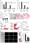
a, Melanoma progression tissue microarray analysis for ABCB5, showing significant differences in ABCB5-staining intensities (mean ± 95% confidence interval (CI); thin or thick nevi versus thin or thick primary melanomas, or versus lymph node or visceral metastases, P values < 0.001; thin versus thick primary melanomas, P = 0.004; thin and thick primary melanomas versus lymph node metastases, P = 0.001; lymph node versus visceral metastases, P = 0.025; n, provided in figure). The picture colour map corresponds to sample types represented in the core array: green, thin nevi; orange, thick nevi; violet, thin primary melanoma; blue, thick primary melanoma; pink, lymph node metastases; yellow, visceral metastases. The scanning view of ABCB5 staining of the entire array corresponds to the colour key. b, Flow cytometry analysis of ABCB5, CD20, nestin, TIE1, CD144, CD31 or BMPR1a expression in n = 7 melanoma patients. c, Marker expression by ABCB5+ or ABCB5 melanoma cells determined by flow cytometry (mean ± s.e.m., n = z4–7 patients).
Figure 2
Figure 2. Tumorigenicity, self-renewal and differentiation of ABCB5+ MMIC
a, Primary tumour formation of unsegregated (US), ABCB5 or ABCB5+ cells. b, Secondary tumour formation of ABCB5 or ABCB5+ cells. c, ABCB5 expression (mean ± s.e.m.) in parent tumours (n = 3) and respective ABCB5+-derived primary (n = 11) and secondary (n = 7) xenografts. d, ABCB5 immunohistochemistry (patient P3). eh, In vivo genetic lineage tracking of human ABCB5+ melanoma cells. e, EYFP versus DsRed plots of a genetically labelled inoculum (left) and a corresponding 6-week-old tumour (right). Controls (small panels): non-transfected cells (top), DsRed+ cells (middle) and EYFP+ cells (bottom). f, Percentage of DsRed+ or EYFP+ cells (mean ± s.e.m.) in inocula (n = 6) and respective tumour xenografts (n = 3) as a function of time. g, Fluorescence microscopy of dissociated 6-week-old xenografts (top and middle rows) and a corresponding frozen tumour section (bottom row). Scale bars, 25 μm. h, DsRed/EYFP positivity in ABCB5+ and ABCB5 6-week-old tumour subpopulations (left); quantified (right) as means ± s.d. (n = 3 replicate experiments).
Figure 3
Figure 3. ABCB5 targeting
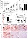
a, Tumour volumes (mean ± s.e.m.) plotted against time. b, tumour formation rate in untreated (n = 18), control-monoclonal-antibody (mAb)-treated (n = 10), or anti-ABCB5 mAb-treated (n = 11) animals. c, Flow cytometric assessment of ADCC in anti-ABCB5 mAb-treated, control mAb-treated or untreated DiO-labelled melanoma target cultures counterstained with propidium iodide (PI). Left panels, representative flow cytometry results showing lysed, DIO+PI+ target cells in the right upper quadrants. Right panel, analysis of ADCC (mean ± s.e.m.) in n = 6 replicate experiments. d, Effect of anti-ABCB5 mAb on established melanoma xenografts. Tumour volumes (mean ± s.e.m.) for anti-ABCB5 mAb-treated (n = 23), control mAb-treated (n = 22), or untreated (n = 22) animals at days 0 and 21 of treatment. e, Immunohistochemistry of patient-derived melanoma xenografts treated with anti-ABCB5 mAb (top) or control mAb (bottom). Adjacent sections were stained with anti-ABCB5 mAb (left), secondary anti-Ig Ab (middle) or CD11b mAb (right), with zones of cellular degeneration in the top row shown below the dotted line.

References

    1. Lapidot T, et al. A cell initiating human acute myeloid leukaemia after transplantation into SCID mice. Nature. 1994;367:645–648. - PubMed
    1. Bonnet D, Dick JE. Human acute myeloid leukemia is organized as a hierarchy that originates from a primitive hematopoietic cell. Nature Med. 1997;3:730–737. - PubMed
    1. Al-Hajj M, et al. Prospective identification of tumorigenic breast cancer cells. Proc Natl Acad Sci USA. 2003;100:3983–3988. - PMC - PubMed
    1. Singh SK, et al. Identification of human brain tumour initiating cells. Nature. 2004;432:396–401. - PubMed
    1. O’Brien CA, Pollett A, Gallinger S, Dick JE. A human colon cancer cell capable of initiating tumour growth in immunodeficient mice. Nature. 2007;445:106–110. - PubMed

Publication types

MeSH terms

Substances