. 2008 Feb;118(2):608-18.
doi: 10.1172/JCI32525.

Factor I is required for the development of membranoproliferative glomerulonephritis in factor H-deficient mice

Affiliations

Factor I is required for the development of membranoproliferative glomerulonephritis in factor H-deficient mice

Kirsten L Rose et al. J Clin Invest. 2008 Feb.

Abstract

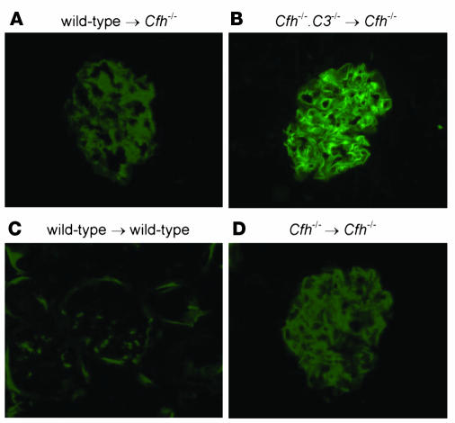
The inflammatory kidney disease membranoproliferative glomerulonephritis type II (MPGN2) is associated with dysregulation of the alternative pathway of complement activation. MPGN2 is characterized by the presence of complement C3 along the glomerular basement membrane (GBM). Spontaneous activation of C3 through the alternative pathway is regulated by 2 plasma proteins, factor H and factor I. Deficiency of either of these regulators results in uncontrolled C3 activation, although the breakdown of activated C3 is dependent on factor I. Deficiency of factor H, but not factor I, is associated with MPGN2 in humans, pigs, and mice. To explain this discordance, mice with single or combined deficiencies of these factors were studied. MPGN2 did not develop in mice with combined factor H and I deficiency or in mice deficient in factor I alone. However, administration of a source of factor I to mice with combined factor H and factor I deficiency triggered both activated C3 fragments in plasma and GBM C3 deposition. Mouse renal transplant studies demonstrated that C3 deposited along the GBM was derived from plasma. Together, these findings provide what we believe to be the first evidence that factor I-mediated generation of activated C3 fragments in the circulation is a critical determinant for the development of MPGN2 associated with factor H deficiency.


Figures

Figure 1
Figure 1. Generation of Cfi–/– mice.
(A) Targeted Cfi locus, targeting vector, and structure of the targeted gene. Exons are represented by filled boxes, and the neomycin resistance gene (Neo) and herpes simplex virus thymidine kinase cassette (HSV-tk) are indicated. Homologous fragments are indicated by dotted lines. E, EcoRI. Double-headed arrows indicate the fragments detected on Southern blot analysis of wild-type and recombinant alleles after EcoRI digestion of genomic DNA hybridized with the 3′ probe (P1, a cDNA probe containing exons 5 and 6). (B) Amplification of recombinant and wild-type alleles using genomic DNA from Cfi–/–, Cfi+/–, and wild-type mice. Absence of the wild-type allele is seen in the Cfi–/– animal. (C) Western blot using nonreducing conditions for serum factor I using crossreactive polyclonal anti-human factor I anti-sera. Lane 1 indicates normal human sera, lane 2 indicates sera from a factor I–deficient individual. Lanes 3–8 represent mouse sera from homozygous, heterozygous, and wild-type mice, as indicated. Absence of the 88-kDa factor I protein (box) is evident in the factor I–deficient individual (lane 2) and in sera from the Cfi–/– mice (lanes 3 and 4).
Figure 2
Figure 2. Complement profile in Cfi–/– mice.
Plasma C3 (A), factor H (B) and factor B (C) levels in Cfi–/–, Cfi+/–, and wild-type mice. Horizontal bars denote median values. Western blot for mouse C3 using EDTA plasma from wild-type and Cfi–/– mice under reducing (D) and nonreducing (E) conditions. In comparison with the molecular weight of the intact C3 α-chain in wild-type plasma, the α-chain of C3 derived from Cfi–/– plasma ran at approximately 110 kDa (D), consistent with the molecular weight of the α′-chain. Under nonreducing conditions C3c was detectable only in EDTA plasma from wild-type mice, as evident by the presence of 130-kDa bands most easily seen on high exposure (E). EDTA plasma dilutions were 1/1000 for both Cfi–/– and wild-type mice.
Figure 3
Figure 3. Light microscopic appearances of mesangial hyalinosis in 8-month-old Cfi–/– mice.
PAS-stained glomerular sections from 8-month-old wild-type (A) and Cfi–/– mice (B and C). The glomerulus from the wild-type mouse had normal appearances. In contrast, areas of mesangial expansion with a nodular hyaline appearance were evident in the Cfi–/– mice (arrows). Original magnification, ×40 (B); ×100(A and C).
Figure 4
Figure 4. Glomerular C3 staining in 8-month-old Cfi–/– mice.
(A) Increased glomerular C3 staining was present in Cfi–/– mice that was mesangial in distribution, in contrast to the linear capillary wall staining pattern seen in Cfh–/– mice (B). Glomerular C3 staining in Cfi+/– mice (C) was identical to that seen in age-matched wild-type animals (D). Quantitative immunofluorescence for glomerular C3 from 8-month-old Cfi–/–, Cfi+/–, and wild-type mice (E). Original magnification, ×40.
Figure 5
Figure 5. Glomerular C3 staining and complement profile in mice with deficiency of both factor H and factor I.
(A) Glomerular C3 staining in Cfi–/– mice with normal (Cfh+/+), heterozygous (Cfh+/–), and homozygous (Cfh–/–) factor H genotypes. A mesangial staining pattern was evident in the glomeruli of the Cfi–/– mice regardless of factor H genotype. Note the marked contrast in glomerular C3 staining pattern between the Cfi–/–Cfh–/– mice and that seen in mice with factor H deficiency alone (Figure 4B). Original magnification, ×40. Plasma C3 (B) and factor B (C) levels in Cfi–/– mice with heterozygous and homozygous factor H deficiency. Horizontal bars denote median values. (D) Western blot for C3 from Cfi–/– and Cfi–/–Cfh–/– mice under reducing conditions. On this high-exposure α-chain, fragments were only present in the Cfh–/– EDTA plasma (lane 4). The lower molecular weight of the α′-chain (lanes 2, 3, and 4) compared with the intact α-chain (lane 1) was also evident. Also shown are sera from Cfi–/– and Cfi–/–Cfh–/– mice before and after incubation with murine sera deficient in C3 (as a source of autologous factors I and H). Note that in both Cfi–/– and Cfi–/–Cfh–/– mice, complete cleavage of the α′-chain occurred with the concomitant appearance of the 43-kDa α′-chain fragment that was evident in unmanipulated EDTA plasma taken from a Cfh–/– animal (lane 4). (E) Under nonreducing conditions C3c was detectable in EDTA plasma from Cfh–/– and wild-type mice, but not from Cfi–/– mice. Note that EDTA plasma dilutions were 1/1000 for wild-type and Cfi–/– mice and 1/100 for Cfh–/– mice.
Figure 6
Figure 6. Administration of sera containing factor I to Cfh–/–Cfi–/– mice.
Following injections of sera from Cfh–/–C3–/– mice (used as a source of autologous factor I), plasma C3 levels fell rapidly to levels comparable with those seen in mice with factor H deficiency alone (A). Furthermore, α′-chain fragments became evident in plasma (B). Finally, at the end of the experiment, florid glomerular C3 staining identical to that seen in unmanipulated Cfh–/– mice was present in the reconstituted animals, in marked contrast to the mesangial C3 staining present in the noninjected Cfh–/–Cfi–/– control mice (C). Consistent with these data was the observation that Cfh–/– mice with heterozygous factor I deficiency had identical glomerular C3 staining to that seen in Cfh–/– animals (D). Original magnification, ×40.
Figure 7
Figure 7. Glomerular C3 staining in renal transplant studies in Cfh–/– mice.
Kidneys from wild-type (A), Cfh–/–C3–/– (B), and Cfh–/– (D) mice were transplanted into Cfh–/– animals. Linear capillary wall C3 staining developed in both wild-type (A) and Cfh–/–C3–/– (B) kidneys transplanted into Cfh–/– animals. Transplantation of a wild-type kidney into a wild-type recipient did not result in abnormal glomerular C3 staining (C), and transplantation of a Cfh–/– kidney into a Cfh–/– recipient did not alter the abnormal glomerular C3 staining seen in unmanipulated Cfh–/– kidneys (D). Original magnification, ×40.

References

    1. Pickering M.C., Botto M., Taylor P.R., Lachmann P.J., Walport M.J. Systemic lupus erythematosus, complement deficiency, and apoptosis. Adv. Immunol. 2000;76:227–324. - PubMed
    1. Liszewski M.K., Farries T.C., Lublin D.M., Rooney I.A., Atkinson J.P. Control of the complement system. Adv. Immunol. 1996;61:201–283. - PubMed
    1. Pangburn M.K., Muller-Eberhard H.J. Relation of putative thioester bond in C3 to activation of the alternative pathway and the binding of C3b to biological targets of complement. J. Exp. Med. 1980;152:1102–1114. - PMC - PubMed
    1. Pangburn M.K., Schreiber R.D., Muller-Eberhard H.J. Formation of the initial C3 convertase of the alternative complement pathway. Acquisition of C3b-like activities by spontaneous hydrolysis of the putative thioester in native C3. . J. Exp. Med. 1981;154:856–867. - PMC - PubMed
    1. Lachmann P.J., Nicol P. Reaction mechanism of the alternative pathway of complement fixation. Lancet. 1973;1:465–467. - PubMed

Publication types

MeSH terms