. 2008 Feb;118(2):597-607.
doi: 10.1172/JCI33263.

IL-22 is required for Th17 cell-mediated pathology in a mouse model of psoriasis-like skin inflammation

Affiliations

IL-22 is required for Th17 cell-mediated pathology in a mouse model of psoriasis-like skin inflammation

Hak-Ling Ma et al. J Clin Invest. 2008 Feb.

Abstract

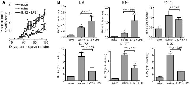
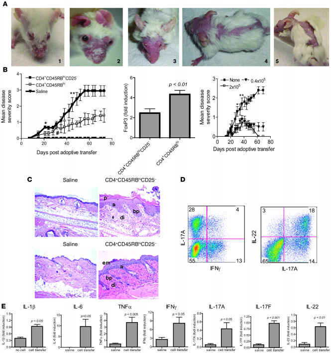
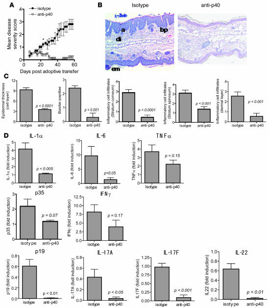
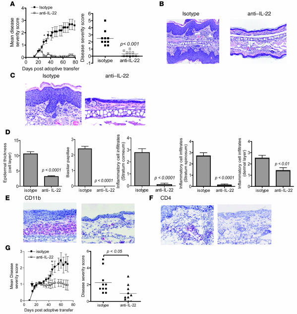
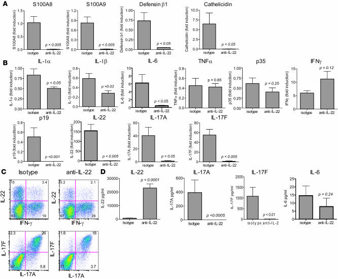
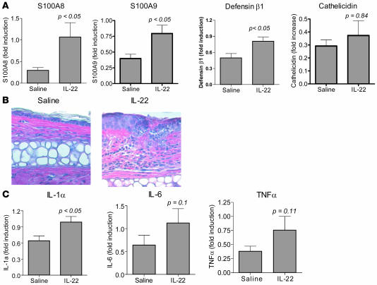
Psoriasis is a chronic skin disease resulting from the dysregulated interplay between keratinocytes and infiltrating immune cells. We report on a psoriasis-like disease model, which is induced by the transfer of CD4(+)CD45RB(hi)CD25(-) cells to pathogen-free scid/scid mice. Psoriasis-like lesions had elevated levels of antimicrobial peptide and proinflammatory cytokine mRNA. Also, similar to psoriasis, disease progression in this model was dependent on the p40 common to IL-12 and IL-23. To investigate the role of IL-22, a Th17 cytokine, in disease progression, mice were treated with IL-22-neutralizing antibodies. Neutralization of IL-22 prevented the development of disease, reducing acanthosis (thickening of the skin), inflammatory infiltrates, and expression of Th17 cytokines. Direct administration of IL-22 into the skin of normal mice induced both antimicrobial peptide and proinflammatory cytokine gene expression. Our data suggest that IL-22, which acts on keratinocytes and other nonhematopoietic cells, is required for development of the autoreactive Th17 cell-dependent disease in this model of skin inflammation. We propose that IL-22 antagonism might be a promising therapy for the treatment of human psoriasis.


Figures

Figure 1
Figure 1. Coadministration of IL-12 and LPS enhances the development of skin lesions in a CD4+CD45RBhi adoptive transfer model.
(A) Disease progression in CB17 scid/scid recipient mice with or without (naive) adoptive transfer of 4 × 10⁵ CD4+CD45RBhi sorted cells. The recipient mice with cell transfer were treated with saline or IL-12 (10 ng) plus LPS (20 μg) (i.p.) on day 1 and saline or IL-12 (10 ng) on day 3 (i.p.). *P < 0.05 starting from day 37 between the saline-treated group and the IL-12/LPS–treated group, both with cell transfer. (B) RNA was purified from an ear biopsy of each mouse within a group at the end of the study and evaluated by quantitative RT-PCR for the indicated cytokines. Results are reported as group means ± SEM, with n = 10 for each group. Data are representative of at least 2 independent experiments. *P < 0.05 between no-treatment group (naive) and saline-treated group with cell transfer; **P < 0.05 between no-treatment group (naive) and LPS plus IL-12–treated group with cell transfer; ***P value between saline and LPS plus IL-12 treated group is indicated.
Figure 2
Figure 2. Adoptive transfer of CD4+CD45RBhi T cells depleted of CD25+ Tregs enhances the development of skin lesions in scid/scid mice.
(A) Representative images of psoriaform lesions in scid/scid mice induced by transfer of CD4+CD45RBhiCD25 T cells. The numbers correspond to the given disease severity scores. (B) Mean disease severity in mice after transfer of indicated cells or saline. Left: *P < 0.05 starting on day 21 between CD4+CD45RBhiCD25 and saline groups; #P < 0.05 starting on day 35 between CD4+CD45RBhi and saline; **P < 0.05 starting on day 49 between CD4+CD45RBhiCD25 and CD4+CD45RBhi (n = 10). Middle: Average Foxp3 gene expression in ear mRNA from indicated groups (n = 10). Right: Mean disease severity in scid/scid mice after transfer of T cells with or without indicated Treg numbers. *P < 0.05 between mice receiving 2 × 10⁵ and no Tregs; **P < 0.05 between 0.4 × 10⁵ and no Tregs. (C) H&E-stained ear (top panels) and back skin (bottom panels) sections from a mouse transferred with saline or CD4+CD45RBhiCD25 cells with parakeratosis (p), acanthosis (a), dermal inflammatory infiltrates (di), basilar papilla (bp), and epidermal microabscess (em) as indicated. Original magnification, ×400. (D) Intracellular cytokine staining was performed on pooled cervical lymph node cells collected from mice that received CD4+CD45RBhiCD25 T cells. Results shown are gated on CD4+ population. (E) Quantitative RT-PCR for the cytokines was performed on ear mRNA, with results reported as group means ± SEM (n = 10). Data are representative of at least 2 independent experiments.
Figure 3
Figure 3. IL-12/IL-23p40 neutralization prevents the development of skin lesions.
(A) Disease severity in CB17 scid/scid mice receiving first CD4+CD45RBhiCD25 cells and subsequently treated with either 0.5 mg of IL-12/23p40 antibody or isotype control antibody (rat IgG2a) i.p. on days 7 and 35 after the adoptive transfer. The mean disease severity scores are reported for 10 mice from each group ± SEM *P < 0.05 starting on day 24, comparing the 2 groups. Data are representative of at least 2 independent experiments. (B) Representative images of H&E-stained sections of ear from recipient CB17 scid/scid mice given an isotype control antibody (left) or IL-12/23p40 antibody (right). Examination of the sections indicates the presence of acanthosis (a), dermal inflammatory infiltrates (di), prominent basilar papilla (bp), and epidermal microabscesses (em) in isotype control antibody–treated mouse skin. Original magnification, ×200. (C) Epidermal thickness and semiquantitative scoring of basilar papillae and inflammatory infiltrates in the stratum corneum, stratum spinosum, and dermal layer of the skin. A section from each mouse ear was scored as follows: 0, within normal limits; 1, minimal; 2, mild; 3, moderate; 4, marked; and 5, severe. The means ± SEM are shown, with n = 10 for each group. (D) Quantitative RT-PCR analysis for each indicated cytokine in RNA isolated from mouse ear at the end of the study. Results reported as mean ± SEM. Data are representative of at least 2 independent experiments, with n = 10 for each group.
Figure 4
Figure 4. IL-22 neutralization prevents the development of skin lesions.
(A) Left panel shows the mean disease severity (±SEM) in recipient mice that were given 16 mg/kg of IL-22 (IL22-104) or isotype control antibody, i.p. once per week for 11 weeks. *P < 0.05 starting on day 26, comparing the 2 groups, with n = 10 mice. Right panel shows the disease severity scores at the end of the study. Data are representative of at least 2 independent experiments. (B and C) H&E-stained sections of ear tissue from recipients given an isotype control (left) or IL-22 antibody (right). Original magnification, ×200 (B) and ×400 (C). (D) Epidermal thickness and semiquantitative scoring of basilar papillae and inflammatory infiltrates in the indicated skin layers. A section from each mouse ear was scored as follows: 0, within normal limits; 1, minimal; 2, mild; 3, moderate; 4, marked; and 5, severe. Means ± SEM are shown, with n = 10 mice. (E and F) Representative immunohistochemical staining for CD11b+ (E) and CD4+ (F) in ear sections from recipients given isotype control antibody (left) or IL-22 antibody (right). Original magnification, ×400. (G) Mean disease severity in recipient mice after cell transfer and subsequent weekly treatment starting on day 14 with 16 mg/kg of IL-22 (IL22-103) or isotype control antibody (as indicated by arrow). *P < 0.05 on day 46 comparing the 2 groups (n = 10 mice). Right panel shows individual disease severity scores at the end of the study.
Figure 5
Figure 5. IL-22 neutralization influences gene expression, T cell phenotype, and cytokine profile.
Recipient mice received an i.p. injection of IL-22 antibody (IL22-104) or isotype control antibody immediately before adoptive transfer and weekly thereafter. At the termination of the study, quantitative RT-PCR was performed on the RNA isolated from individual mouse ears for transcripts encoding (A) antimicrobial peptides and (B) cytokines. Results are reported as means ± SEM. All data are representative of at least 2 independent experiments, with n = 10 for each group. (C) Intracellular cytokine staining for IL-22, IL-17A, IL-17F, and IFN-γ performed on pooled cervical lymph node cells collected from mice treated with isotype control or IL-22 antibody. Results shown are gated on the CD4+ population. Data are representative of at least 2 independent experiments. (D) Serum was collected at the termination of a study from mice given isotype or IL-22 antibody. Serum IL-22, IL-17A, IL-17F, and IL-6 were determined by standard ELISA. Results are reported as group means ± SEM, with n = 10 for each group. TNF-α and IFN-γ were below the limit of detection of this assay. Data are representative of at least 2 independent experiments.
Figure 6
Figure 6. IL-22 induces antimicrobial peptide and proinflammatory cytokine gene expression and keratinocyte hyperplasia in the skin.
Ears of BALB/c mice were injected intradermally every other day for 2 weeks with 500 ng of IL-22 or saline in a total volume of 20 μl. (A) 6 hours after the first injection, mouse ears were harvested, RNA purified, and antimicrobial peptide transcript levels evaluated by quantitative RT-PCR. Results are reported as group means ± SEM, with n = 5 for each group. (B) H&E-stained sections 2 weeks after injection from ears injected with saline (left panel) or IL-22 (right panel). Note the keratinocyte proliferation and inflammatory infiltrates. Original magnification, ×400. (C) After 2 weeks of injections, mouse ears were harvested, RNA purified, and cytokine transcript levels evaluated by quantitative RT-PCR. Results are reported as group means ± SEM, with n = 5 for each group. Data are representative of at least 2 independent experiments.

References

    1. Schon M.P., Boehncke W.H. Psoriasis. N. Engl. J. Med. 2005;352:1899–1912. - PubMed
    1. Zenz R., et al. Psoriasis-like skin disease and arthritis caused by inducible epidermal deletion of Jun proteins. Nature. 2005;437:369–375. - PubMed
    1. Cook P.W., Brown J.R., Cornell K.A., Pittelkow M.R. Suprabasal expression of human amphiregulin in the epidermis of transgenic mice induces a severe, early-onset, psoriasis-like skin pathology: Expression of amphiregulin in the basal epidermis is also associated with synovitis. Exp. Dermatol. 2004;13:347–356. - PubMed
    1. Lowes M.A., Bowcock A.M., Krueger J.G. Pathogenesis and therapy of psoriasis. Nature. 2007;445:866–873. - PubMed
    1. Groves R.W., et al. Inflammatory and hyperproliferative skin disease in mice that express elevated levels of the IL-1 receptor (Type I) on epidermal keratinocytes. Evidence that IL-1-inducible secondary cytokines produced by keratinocytes in vivo can cause skin disease. J. Clin. Invest. 1996;98:336–344. - PMC - PubMed

MeSH terms