Comparative Study
. 2008 Feb;38(2):459-69.
doi: 10.1002/eji.200737268.

Mannose-capped lipoarabinomannan- and prostaglandin E2-dependent expansion of regulatory T cells in human Mycobacterium tuberculosis infection

Affiliations
Comparative Study

Mannose-capped lipoarabinomannan- and prostaglandin E2-dependent expansion of regulatory T cells in human Mycobacterium tuberculosis infection

Ankita Garg et al. Eur J Immunol. 2008 Feb.

Abstract

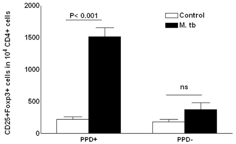
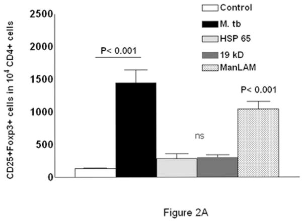
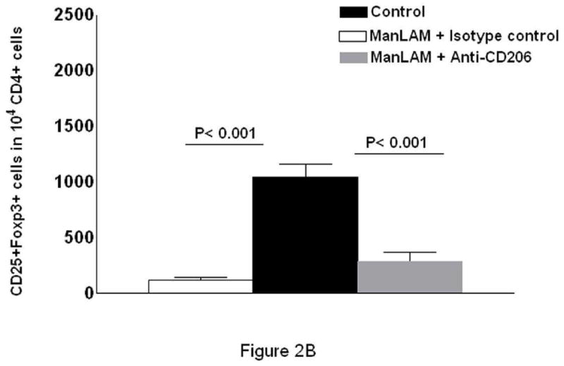
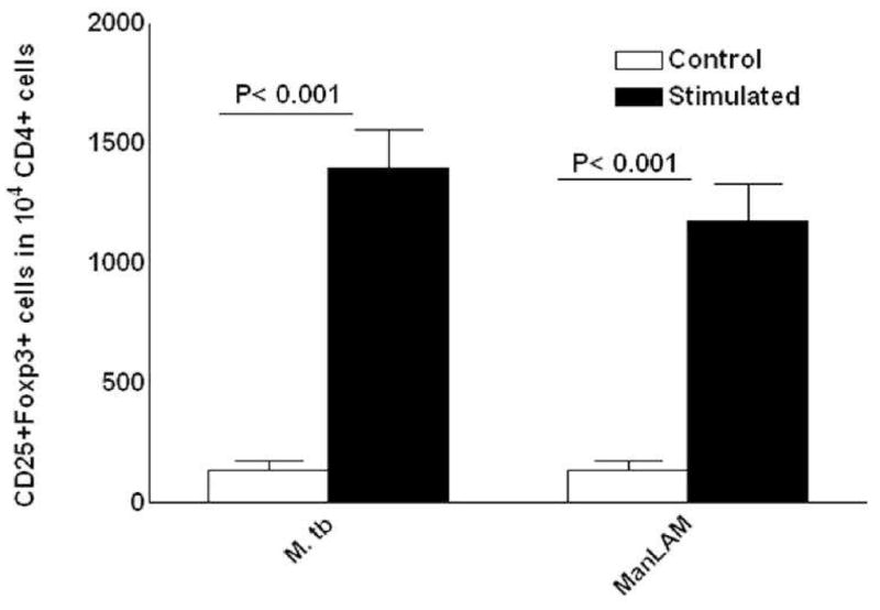
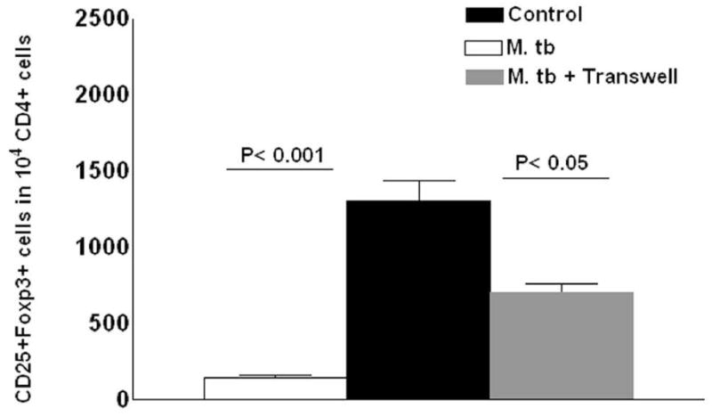
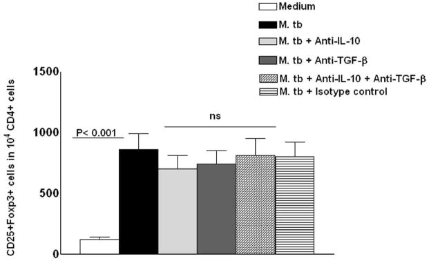
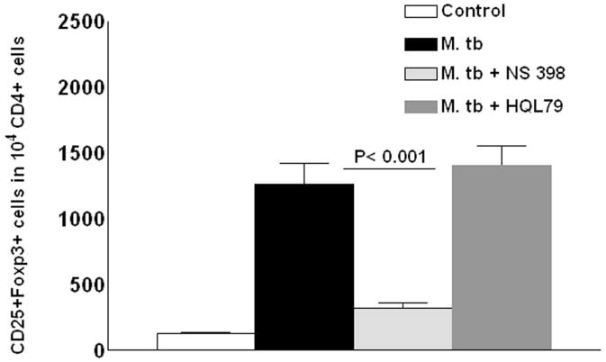
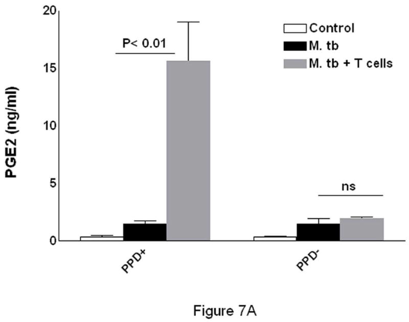
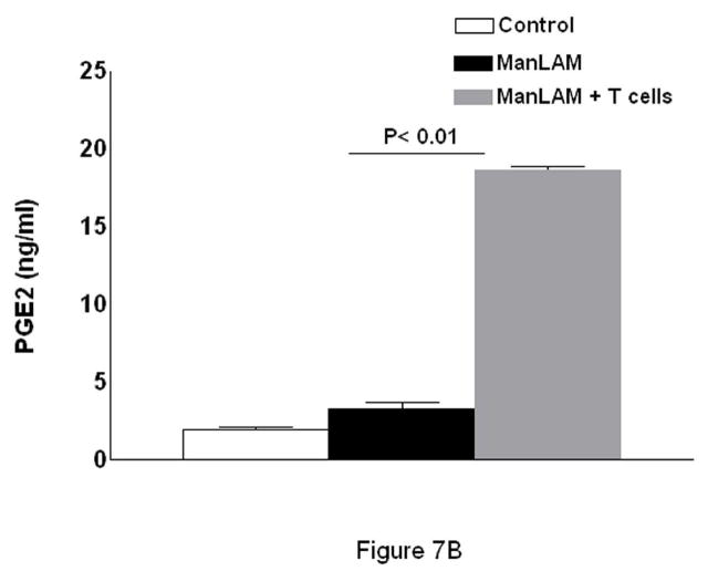
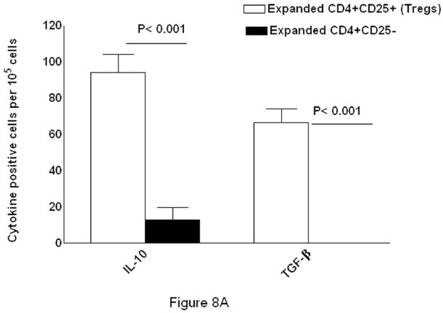
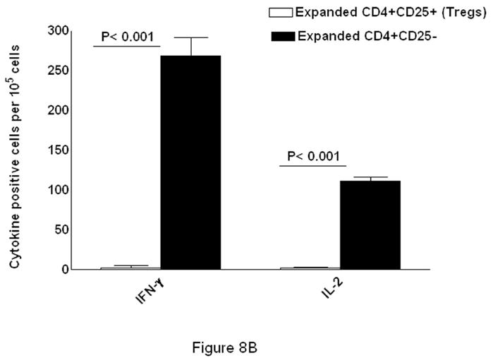
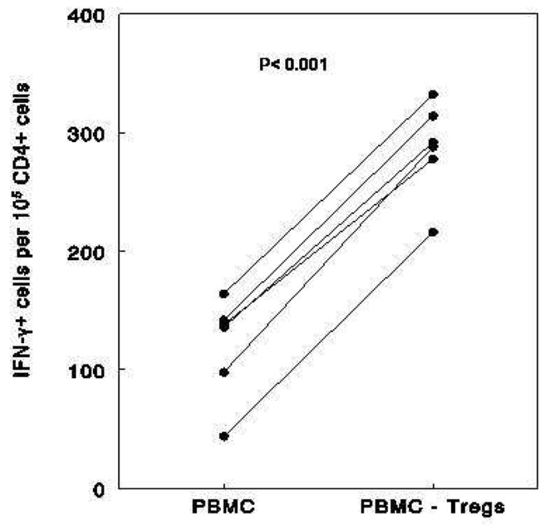
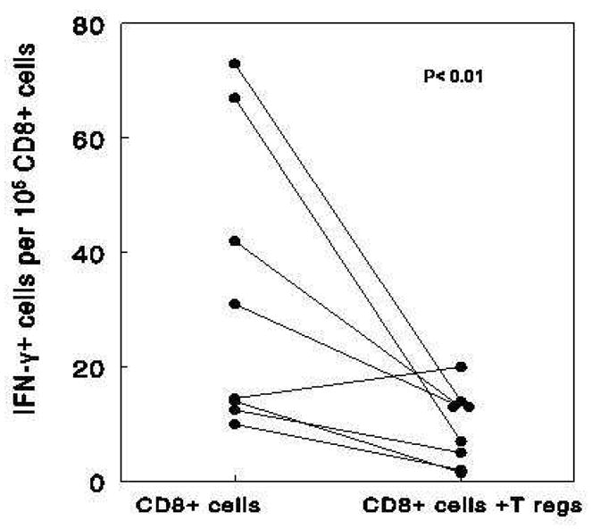
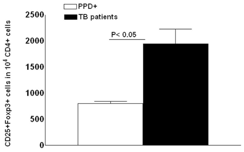
We evaluated the role of regulatory T cells (CD4(+) CD25(+) Foxp3(+) cells, Tregs) in human Mycobacterium tuberculosis infection. Tregs were expanded in response to M. tuberculosis in healthy tuberculin reactors, but not in tuberculin-negative individuals. The M. tuberculosis mannose-capped lipoarabinomannan (ManLAM) resulted in regulatory T cell expansion, whereas the M. tuberculosis 19-kDa protein and heat shock protein 65 had no effect. Anti-IL-10 and anti-TGF-beta alone or in combination, did not reduce expansion of Tregs. In contrast, the cyclooxygenase enzyme-2 inhibitor NS398 significantly inhibited expansion of Tregs, indicating that prostaglandin E2 (PGE2) contributes to Treg expansion. Monocytes produced PGE2 upon culturing with heat-killed M. tuberculosis or ManLAM, and T cells from healthy tuberculin reactors enhanced PGE2 production by monocytes. Expanded Tregs produced significant amounts of TGF-beta and IL-10 and depletion of Tregs from PBMC of these individuals increased the frequency of M. tuberculosis-responsive CD4(+) IFN-gamma cells. Culturing M. tuberculosis-expanded Tregs with autologous CD8(+) cells decreased the frequency of IFN-gamma(+)cells. Freshly isolated PBMC from tuberculosis patients had increased percentages of Tregs, compared to healthy tuberculin reactors. These findings demonstrate that Tregs expand in response to M. tuberculosis through mechanisms that depend on ManLAM and PGE2.


Conflict of interest statement

The authors have no financial conflict of interest.

Figures

Figure 1
Figure 1. Expansion of Tregs by monocytes exposed to M. tuberculosis
Effect of prior M. tuberculosis infection. Freshly isolated CD4+ cells and autologous monocytes from 14 healthy tuberculin reactors (PPD+) and four healthy tuberculin-negative subjects (PPD-) were cultured at a ratio of 9:1, in the presence or absence of heat-killed M. tuberculosis Erdman (M. tb, 10 μg/ml). After 5 days, the percentages of CD4+CD25+Foxp3+ cells were determined by flow cytometry, and expressed as the number of CD4+CD25+ cells per 104 CD4+cells. Mean values and SEs are shown.
Figure 2
Figure 2. Effect of ManLAM on expansion of Tregs
Panel A. ManLAM-mediated expansion of Tregs. CD4+ cells from six healthy tuberculin reactors were cultured with autologous monocytes in medium alone, or with HSP65, the hemaxeric peptide of the 19-kD M. tuberculosis lipoprotein, or ManLAM (all at 10 μg/ml). After 5 days, the percentages of CD4+CD25+Foxp3+ cells were determined by flow cytometry and expressed as the number of CD4+CD25+ cells per 104 CD4+ cells. Mean values and SEs are shown. p< 0.001. Panel B. Effect of blocking the mannose receptor on expansion of Tregs. CD4+ cells from seven healthy tuberculin reactors were cultured with autologous monocytes in the presence of medium alone, or with ManLAM and anti-CD206 (10 μg/ml) or isotype control Ab (10 μg/ml). After 5 days, the percentages of CD4+CD25+Foxp3+ cells were determined by flow cytometry, and expressed as the number of CD4+CD25+ cells per 104 CD4+ cells. Mean values and SEs are shown.
Figure 3
Figure 3. M. tuberculosis and ManLAM converts some CD4+CD25-Foxp3- cells to CD4+CD25+Foxp3+ cells
CD4+ cells from five healthy tuberculin reactors were isolated and CD25+ cells were removed, using the Tregs isolation kit, as described in the methods section. The remaining CD4+CD25- cells were cultured with autologous monocytes in medium alone, or with M. tuberculosis whole cell lysate, or ManLAM (each at 10 μg/ml). After 5 days, the percentages of CD4+CD25+Foxp3+ cells were determined by flow cytometry and the results were expressed as the number of CD4+CD25+ cells per 104 CD4+ cells. Mean values and SEs are shown.
Figure 4
Figure 4. Effect of soluble factors produced by monocytes on expansion of Tregs
Purified CD4+ cells from four healthy tuberculin reactors were cultured in 12-well plates. Transwell inserts in some wells contained monocytes, with or without heat-killed M. tuberculosis. In other wells, M. tuberculosis–exposed monocytes were cultured with CD4+ cells in the same well. After 5 days, the percentages of CD4+CD25+Foxp3+ cells were determined by flow cytometry and the results were expressed as the number of CD4+CD25+ cells per 104 CD4+ cells. Mean values and SEs are shown.
Figure 5
Figure 5. Effect of neutralization of IL-10 and TGF-β on expansion of Tregs
CD4+ cells and autologous monocytes from five healthy tuberculin reactors were cultured in medium alone or with heat-killed M. tuberculosis. Anti-IL-10 (10 μg/ml) and anti-TGF-β (10 μg/ml), either alone or together (5 μg/ml each), or an isotype control Ab (10 μg/ml), were added to some M. tuberculosis-stimulated wells. After 5 days, the percentages of CD4+CD25+Foxp3+ cells were determined by flow cytometry, and the results were expressed as the number of CD4+CD25+ cells per 104 CD4+ cells. Mean values and SEs are shown.
Figure 6
Figure 6. Effect of PGE2 inhibition on expansion of Tregs
Freshly isolated CD4+ cells and autologous monocytes from 7 healthy tuberculin reactors were cultured with heat-killed M. tuberculosis. NS398, which inhibits formation of PGE2, or HQL79, which inhibits formation of PGD2, were added to some M. tuberculosis–stimulated wells. After 5 days, the percentages of CD4+CD25+Foxp3+ cells were determined by flow cytometry, and the results were expressed as the number of CD4+CD25+ cells per 104 CD4+ cells. Mean values and SEs are shown.
Figure 7
Figure 7. Production of PGE2 by monocytes stimulated with M. tuberculosis (panel A) or ManLAM (panel B)
A. Freshly isolated monocytes from 5 healthy tuberculin reactors and 4 healthy tuberculin-negative donors were cultured with or without 10 μg/ml of heat-killed M. tuberculosis Erdman. To some M. tuberculosis-stimulated monocytes, CD4+ cells were added at a ratio of 1 T-cell to 4 monocytes. After 72 h, PGE2 concentrations in supernatants were measured by ELISA. Mean values ± SEs are shown. B. Freshly isolated monocytes from 6 healthy tuberculin reactors were cultured with or without 10 μg/ml of ManLAM. To some ManLAM-stimulated monocytes, CD4+ cells were added at a ratio of 1 T-cell to 4 monocytes. After 72 h, PGE2 concentrations in supernatants were measured by ELISA. Mean values ± SEs are shown.
Figure 8
Figure 8. Cytokine production by expanded CD4+CD25+ (Tregs) and CD4+CD25- cells
Panel A. TGF-β and IL-10 production. CD4+ cells and autologous monocytes from six healthy tuberculin reactors were cultured in medium alone or with M. tuberculosis whole cell lysate. After 48 h, CD4+ CD25+ cells (Tregs) and CD4+ CD25- cells were isolated, as described in the methods section, and incubated overnight in triplicate wells on an ELISPOT plate to detect TGF-β- and IL-10-producing cells. Mean values ± SEs are shown. Panel B. IFN- γ and IL-2 production. CD4+ cells and autologous monocytes from six healthy tuberculin reactors were cultured, as in panel A. After isolation, CD4+ CD25+ cells (Tregs) and CD4+ CD25- cells were incubated overnight in triplicate wells on an ELISPOT plate to detect IFN-γ and IL-2-producing cells. Mean values ± SEs are shown.
Figure 9
Figure 9. Tregs reduce the frequency of M. tuberculosis-responsive IFN-γ + T cells
Panel A. Effect of depletion of Tregs on the frequency of M. tuberculosis-responsive CD4+ IFN-γ + T cells. PBMC and Treg-depleted PBMC were obtained from 5 healthy tuberculin reactors. Treg-depleted PBMC were obtained by positively selecting CD4+ CD25+ cells from PBMC, using the Treg isolation kit, followed by pooling all remaining CD4- and CD4+CD25- fractions. PBMC and Treg-depleted PBMC were cultured with M. tuberculosis for 48 h. CD4+ cells were isolated from PBMC and from Treg-depleted PBMC and incubated overnight in triplicate wells on an ELISPOT plate to detect IFN-γ-producing cells. Panel B. M. tuberculosis-expanded Tregs reduce the frequency of M. tuberculosis-responsive CD8+IFN-γ + T cells. Freshly isolated CD4+ cells and autologous monocytes from 8 healthy tuberculin reactors were cultured with heat-killed M. tuberculosis. After 72 h, Tregs were isolated and cultured with freshly isolated autologous CD8+ cells, and CD14+ cells with heat-killed M. tuberculosis at a ratio of 1:9:1. After 2 more days, CD8+ cells were isolated and incubated overnight in triplicate wells on an ELISPOT plate to detect IFN-γ-producing cells.
Figure 10
Figure 10. Tregs in healthy tuberculin reactors and tuberculosis patients
Freshly isolated PBMC from 6 healthy tuberculin reactors and 9 tuberculosis patients were stained with Abs to CD4, CD25 and Foxp3. Flow cytometry was used to measure CD4+CD25+Foxp3+ cells, and the results were expressed as the number of CD4+CD25+ cells per 104 CD4+ cells. Mean values and standard errors are shown.

Similar articles

Cited by

References

    1. Baumann S, Eddine AN, Kaufmann SH. Progress in tuberculosis vaccine development. Curr Opin Immunol. 2006;18:438–448. - PubMed
    1. Horsburgh CR., Jr Priorities for the treatment of latent tuberculosis infection in the United States. N Engl J Med. 2004;350:2060–2067. - PubMed
    1. Rojas RE, Balaji KN, Subramanian A, Boom WH. Regulation of human CD4(+) alphabeta T-cell-receptor-positive (TCR(+)) and gammadelta TCR(+) T-cell responses to Mycobacterium tuberculosis by interleukin-10 and transforming growth factor beta. Infect Immun. 1999;67:6461–6472. - PMC - PubMed
    1. Cooper AM, Dalton DK, Stewart TA, Griffin JP, Russell DG, Orme IM. Disseminated tuberculosis in interferon gamma gene-disrupted mice. J Exp Med. 1993;178:2243–2247. - PMC - PubMed
    1. Flynn JL, Chan J, Triebold KJ, Dalton DK, Stewart TA, Bloom BR. An essential role for interferon gamma in resistance to Mycobacterium tuberculosis infection. J Exp Med. 1993;178:2249–2254. - PMC - PubMed

Publication types

MeSH terms