Review
. 2007;2(4):515-26.

Clinical applications of perfluorocarbon nanoparticles for molecular imaging and targeted therapeutics

Affiliations
Review

Clinical applications of perfluorocarbon nanoparticles for molecular imaging and targeted therapeutics

Trung D Tran et al. Int J Nanomedicine. 2007.

Abstract

Molecular imaging is a novel tool that has allowed non-invasive diagnostic imaging to transition from gross anatomical description to identification of specific tissue epitopes and observation of biological processes at the cellular level. This technique has been confined to the field of nuclear imaging; however, recent advances in nanotechnology have extended this research to include ultrasound (US) and magnetic resonance (MR) imaging. The exploitation of nanotechnology for MR and US molecular imaging has generated several candidate contrast agents. One multimodality platform, targeted perfluorocarbon (PFC) nanoparticles, is useful for noninvasive detection with US and MR, targeted drug delivery, and quantification.


Figures

Figure 1
Figure 1
Scanning electron micrographs (×30 000) of control fibrin clot (A) and fibrin-targeted paramagnetic nanoparticles bound to the clot surface (B). Arrows indicate (A) fibrin fibril; (B) fibrin-specific nanoparticles-bound to fibrin epitopes. Copyright © 2001. Reprinted with permission from Flacke S, Fischer S, et al. 2001. Novel MRIM contrast agent for molecular imaging of fibrin: implications for detecting vulnerable plaques. Circulation, 104:1280–5.
Figure 2
Figure 2
Paradigm for targeted liquid perfluorocarbon-based nanoparticle contrast agents. This example has a payload of Gd3+ chelates and monoclonal antibodies. This platform is extremely versatile and applicable to almost any imaging modalities and capable of carrying other payloads such as drugs or genes.
Figure 3
Figure 3
(Left) Low-resolution images (3D gradient and spin echo) of control and (Middle) fibrin-targeted clot with paramagnetic nanoparticles presenting a homogenous, T1-weighted enhancement. (Right) High resolution scans of fibrin clots (3D T1-weighted gradient recalled echo sequence), revealing that image B results from a thin layer of paramagnetic nanoparticles along the surface. Copyright © 2001. Reprinted with permission from Flacke S, Fischer S, et al. 2001. Novel MRI contrast agent for molecular imaging of fibrin: implications for detecting vulnerable plaques. Circulation, 104:1280–5.
Figure 4
Figure 4
Color enhanced magnetic resonance imaging of fibrin-targeted and control carotid endarterectomy speciments revealing contrast enhancement (white) of a small fibrin deposit on a symptomatic ruptured plaque. Calcium deposit (black). 3D, fat-suppressed, T1-weighted fast gradient echo. Copyright © 2001. Reprinted with permission from Flacke S, Fischer S, et al. 2001. Novel MRI contrast agent for molecular imaging of fibrin: implications for detecting vulnerable plaques. Circulation, 104:1280–5.
Figure 5
Figure 5
(a) Representative spectrum taken at 4.7 T of crown ether emulsion (~90 ppm) and trichlorofluormethane (0 ppm) used as a reference. (b) The calibration curve for crown ether emulsion has a slope of 28.06 with an r2 of 0.9968. (c) The calculated number of bound nanoparticles (mean ± standard error, n = 3) as calculated from 19F spectroscopy versus the mass of total gadolinium (Gd3+) in the sample as determined by neutron activation analysis show excellent agreement as independent measures of fibrin-targeted nanoparticles binding to clots. The linear regression line has an r2 of 0.9997. Copyright © 2004. Reprinted with permission from Morawski AM, Winter PM, et al. 2004. Targeted nanoparticles for quantitative imaging of sparse molecular epitopes with MRI. Magnetic Resonance in Medicine, 51:480–6.
Figure 6
Figure 6
(a) Optical image of a 5-mm cross-section of a human carotid endarterectomy sample. This section showed moderate luminal narrowing as well as several atherosclerotic lesions. (b) A 19F projection image acquired at 4.7 T through the entire carotid artery sample shows high signal along the lumen due to nanoparticles bound to fibrin. (c) Concentration map of bound nanoparticles in the carotid sample. Copyright © 2004. Reprinted with permission from Morawski AM, Winter PM, et al. 2004. Targeted nanoparticles for quantitative imaging of sparse molecular epitopes with MRI. Magnetic Resonance in Medicine, 51:480–6.
Figure 7
Figure 7
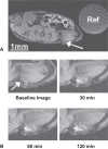
(A) T1-weighted MR image (axial view) of an athymic nude mouse before injection of paramagnetic ανβ3-targeted nanoparticles. Arrow indicates a C32 tumor that is difficult to detect (“Ref” = Gd3+ DTPA in 10 cc syringe). (B) Enlarged section of an MR image showing T1-weighted signal enhancement of angiogenic vasculature of early tumors over 2 h as detected by ανβ3-targeted nanoparticles. Copyright © 2005. Reprinted with permission from Schmieder AH, Winter PM, et al. 2005. Molecular MR imaging of melanoma angiogenesis with alphanubeta3-targeted paramagnetic nanoparticles. Magnetic Resonance in Medicine, 53:621–7.
Figure 8
Figure 8
In vivo spin-echo image reformatted to display long axis of aorta from renal arteries to diaphragm of a cholesterol-fed rabbit (top) and at single transverse level (bottom) before (Pre) and after (Post) treatment, after semi-automated segmentation (Segmented, grayish ring), and with color-coded signal enhancement (Enhancement) above baseline (in percent). Copyright © 2003. Reprinted with permission from Winter PM, Morawski AM, et al. 2003. Molecular imaging of angiogenesis in early-stage atherosclerosis with alpha(v)beta3-integrin-targeted nanoparticles. Circulation, 108:2270–4.
Figure 9
Figure 9
Schematic representation illustrating contact-facilitated drug delivery. Phospholipids and drug within the nanoparticles surfactant exchange with lipids of the target membrane through a convection process, rather than diffusion, as is common among other targeted systems. Copyright © 2004. Reprinted with permission from Lanza GM, Winter PM, et al. 2004. Magnetic resonance molecular imaging with nanoparticles. Journal of Nuclear Cardiology, 11:733–43.
Figure 10
Figure 10
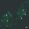
In vitro targeting of FITC-labeled nanoparticles (white arrows) targeted to aνb3-integrin expressed by C-32 melanoma cells, which illustrate the delivery of FITC-labeled surfactant lipids into target cell membranes (yellow arrows). Scale bar = 2 microns. Copyright © 2004. Reprinted with permission from Lanza GM, Winter PM, et al. 2004. Magnetic resonance molecular imaging with nanoparticles. Journal of Nuclear Cardiology, 11:733–43.

References

    1. Ambrose JA, Tannenbaum MA, et al. Angiographic progression of coronary artery disease and the development of myocardial infarction. Journal of the American College of Cardiology. 1988;12:56–62. - PubMed
    1. Anderson SA, Rader RK, et al. Magnetic resonance contrast enhancement of neovasculature with alpha(v)beta(3)-targeted nanoparticles. Magnetic Resonance in Medicine. 2000;44:433–9. - PubMed
    1. Bishop GG, McPherson JA, et al. Selective alpha(v)beta(3)-receptor blockade reduces macrophage infiltration and restenosis after balloon angioplasty in the atherosclerotic rabbit. Circulation. 2001;103:1906–11. - PubMed
    1. Blomley MJ, Cooke JC, et al. Microbubble contrast agents: a new era in ultrasound. BMJ. 2001;322:1222–5. - PMC - PubMed
    1. Brooks PC, Clark RA, et al. Requirement of vascular integrin alpha v beta 3 for angiogenesis. Science. 1994;264:569–71. - PubMed

MeSH terms