

Complete anterior cruciate ligament tear and the risk for cartilage loss and progression of symptoms in men and women with knee osteoarthritis
- PMID: 18203629
- PMCID: PMC3125710
- DOI: 10.1016/j.joca.2007.11.005
Complete anterior cruciate ligament tear and the risk for cartilage loss and progression of symptoms in men and women with knee osteoarthritis
Abstract
Objective: To determine whether a complete anterior cruciate ligament (ACL) tear, a frequent incidental finding on magnetic resonance imagings (MRIs) of individuals with established knee osteoarthritis (OA), increases the risk for further knee OA progression.
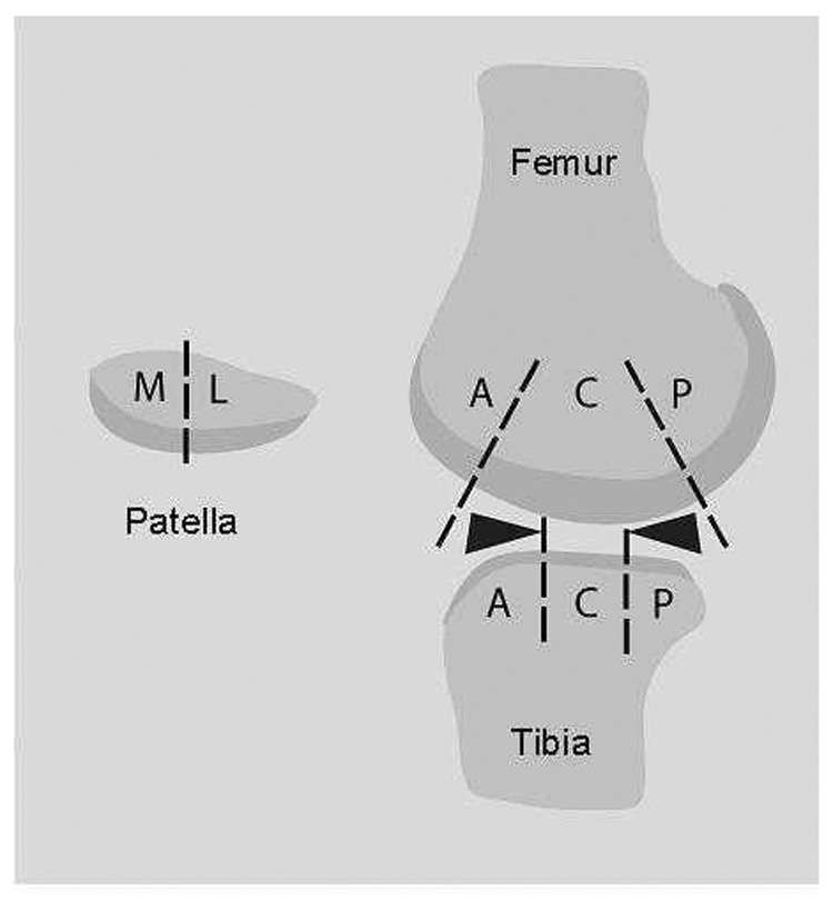
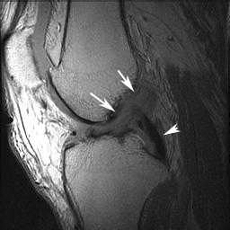
Methods: We examined 265 participants (43% women) with symptomatic knee OA in a 30-month, prospective, natural history study of knee OA. The more symptomatic knee was imaged using MRI at baseline, 15 and 30 months. Cartilage was scored at the medial and lateral tibiofemoral joint and at the patellofemoral joint using the Whole-Organ MRI Score (WORMS) semi-quantitative method. Complete ACL tear was determined on baseline MRI. At each visit, knee pain was assessed using a knee-specific visual analog scale and physical function was assessed using the Western Ontario and McMaster Universities (WOMAC) physical function subscale.
Results: There were 49 participants (19%) with complete ACL tear at baseline. Adjusting for age, body mass index, gender and baseline cartilage scores, complete ACL tear increased the risk for cartilage loss at the medial tibiofemoral compartment [odds ratio (OR): 1.8, 95% confidence interval (CI): 1.1, 3.2]. However, following adjustment for the presence of medial meniscal tears, no increased risk for cartilage loss was further seen (OR: 1.1, 95% CI: 0.6, 1.8). Knee pain and physical function were similar over follow-up between those with and without a complete ACL tear.
Conclusions: Individuals with knee OA and incidental complete ACL tear have an increased risk for cartilage loss that appears to be mediated by concurrent meniscal pathology. The presence of a complete ACL tear did not influence the level of knee pain or physical function over short-term follow-up.
Figures
References
-
- Hill CL, Seo GS, Gale D, Totterman S, Gale ME, Felson DT. Cruciate ligament integrity in osteoarthritis of the knee. Arthritis & Rheumatism. 2005;52(3):794–799. - PubMed
-
- Link TM, Steinbach LS, Ghosh S, Ries M, Lu Y, Lane N, et al. Osteoarthritis: MR imaging findings in different stages of disease and correlation with clinical findings. Radiology. 2003;226(2):373–381. - PubMed
-
- Chan WP, Lang P, Stevens MP, Sack K, Majumdar S, Stoller DW, et al. Osteoarthritis of the knee: comparison of radiography, CT, and MR imaging to assess extent and severity. AJR. American Journal of Roentgenology. 1991;157(4):799–806. - PubMed
-
- Cushner FD, La Rosa DF, Vigorita VJ, Scuderi GR, Scott WN, Insall JN. A quantitative histologic comparison: ACL degeneration in the osteoarthritic knee. Journal of Arthroplasty. 2003;18(6):687–692. - PubMed
-
- Chan WP, Peterfy C, Fritz RC, Genant HK. MR diagnosis of complete tears of the anterior cruciate ligament of the knee: importance of anterior subluxation of the tibia. AJR. American Journal of Roentgenology. 1994;162(2):355–360. - PubMed
Publication types
MeSH terms
Grants and funding
LinkOut - more resources
Full Text Sources
Other Literature Sources
