. 2008 Jan 29;105(4):1164-9.
doi: 10.1073/pnas.0702132105. Epub 2008 Jan 18.

BiP mutants that are unable to interact with endoplasmic reticulum DnaJ proteins provide insights into interdomain interactions in BiP

Affiliations

BiP mutants that are unable to interact with endoplasmic reticulum DnaJ proteins provide insights into interdomain interactions in BiP

Walid Awad et al. Proc Natl Acad Sci U S A. .

Abstract

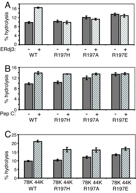
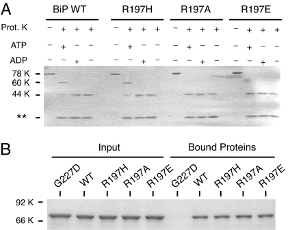
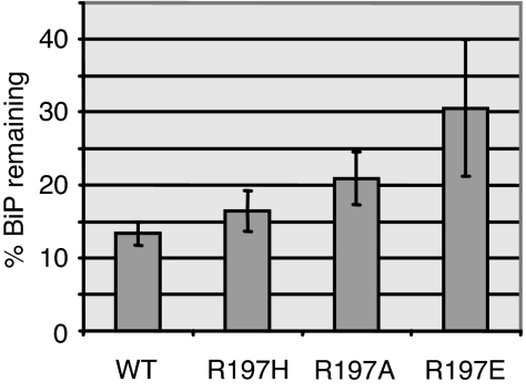
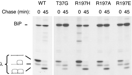
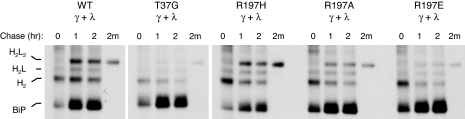
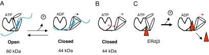
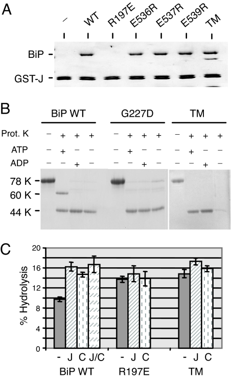
The heat shock protein (Hsp)70 family of molecular chaperones interacts with unfolded proteins through a C-terminal substrate-binding domain (SBD) that is controlled by nucleotide binding to the N-terminal domain. The ATPase cycle is regulated by cochaperones, including DnaJ proteins that accelerate ATP hydrolysis to stabilize the Hsp70-substrate complex. We found that R197 in hamster BiP, which resides at the surface of the nucleotide-binding domain, is critical for both association with endoplasmic reticulum DnaJ proteins and interaction with the SBD. Decreasing the positive charge at this residue enhanced basal ATPase activity, destabilized interaction with the SBD, and reduced substrate release both in vitro and in vivo. Mutation of three glutamic acids in the SBD mimicked many of these effects. Our data provide insights into communications between the two domains and suggest a mechanism by which DnaJ proteins increase ATP hydrolysis.


Conflict of interest statement

The authors declare no conflict of interest.

Figures

Fig. 1.
Fig. 1.
Characterization of BiP R197 mutants. (A) Recombinant protein was isolated for full-length and N-terminal nucleotide-binding fragment of wild-type BiP and three different mutants with amino acid substitutions at residue 197. Full-length recombinant BiP proteins were assayed for ATPase activity when stimulated with full-length recombinant ERdj3 (A) or peptide C (B). (C) The basal ATPase activity of the full-length and NBD of wild-type and mutant proteins was measured.
Fig. 2.
Fig. 2.
Mutations at residue 197 do not affect nucleotide binding but do alter nucleotide-induced conformational changes. (A) Recombinant BiP proteins were incubated in the absence of proteinase K or with proteinase K, along with ATP, ADP, or nucleotide. Samples were analyzed by SDS/PAGE. Full-length proteins migrated at ≈78 kDa, whereas the normal ATP-protected fragment migrated at ≈60 kDa, and the ADP-protected fragment migrated at ≈44 kDa. Proteinase K can be observed at the bottom of the gel. (B) The three R197 mutants, as well as wild-type and G227D BiP, were incubated with ATP–agarose to assess the effects of the mutations on nucleotide binding. The left side of the gel is the input protein, and right side is the protein bound to the ATP–agarose beads. G227D BiP serves as a control for a non-ATP-binding mutant.
Fig. 3.
Fig. 3.
ATP-mediated release of R197 mutants from substrate in vitro. COS cells were cotransfected with Ig HCs and either wild-type BiP or the three R197 mutants. Cells were metabolically labeled, and lysates were divided for immunoprecipitation with anti-BiP or protein A–Sepharose to isolate Ig HCs. One set of HCs was left untreated, and one set was incubated with ATP for each group. Samples were electrophoresed on 10% SDS gels under reducing conditions. BiP signals were quantitated by phosphorimaging, normalized to HC signals, and expressed as the percentage remaining after ATP treatment. Results are from three independent experiments, and error bars represent SD.
Fig. 4.
Fig. 4.
R197 substitutions can affect Ig light chain folding. COS cells were transfected with the indicated BiP constructs along with mouse λ light chains. Cells were pulse-labeled with [35S]methionine in the presence of DTT (0) and then chased in complete media without DTT for 45 min. Cell lysates were immunoprecipitated with anti-λ antibodies, and proteins were electrophoresed under nonreducing conditions. The migration of completely reduced, partially oxidized, and completely oxidized light chains is indicated at the right.
Fig. 5.
Fig. 5.
Effects of R197 substitutions on Ig assembly. COS cells were transfected with the indicated BiP constructs along with vectors encoding Ig γ HC and λ light chain. Cells were pulse-labeled (0) and chased for the indicated times. The media from the 2-h chase (2m) was saved, and both cell lysates and culture supernatants were immunoprecipitated with anti-γ HC antiserum. Samples were analyzed under nonreducing conditions, and the mobility of assembly intermediates is indicated at the left.
Fig. 6.
Fig. 6.
Model for functional interactions between nucleotide and substrate-binding domains. (A) When BiP is in the ATP-bound form, which is open for substrate binding, the SBD interacts with the NBD in a way resulting in the protection of a 60-kDa fragment when the protein is treated with protease. Hydrolysis of ATP induces closing of the SBD and an alteration in its interaction of the NBD, so that only a 44-kDa fragment is now protected. (B) Mutation of R197 (red asterisk) on the NBD to a neutral or negatively charged residue mimics the ADP-bound form in that the two domains no longer interact in a way that allows protection of a 60-kDa fragment and induces closing of the SBD. (C) ERdj3 (triangle) binds to the ATP-bound form of BiP and serves to push the SBD domain away from the NBD and thus relieve the suppression to ATP hydrolysis that this domain provides and concomitantly to close the SBD.
Fig. 7.
Fig. 7.
Mutation of negative residues on the SBD of BiP can affect interdomain communication and ATP hydrolysis. (A) Recombinant proteins corresponding to wild-type BiP, three single-point mutants in the SBD, and a TM in which all three residues were combined were incubated with a fusion protein corresponding to ERdj3 J domain and GST. Bound material was subjected to SDS/PAGE analysis and detected by Coomassie blue staining. (B) The indicated recombinant BiP proteins were incubated with ATP, ADP, or no nucleotide and treated with proteinase K as described in Fig. 2. (C) Recombinant BiP proteins were analyzed for ATPase activity alone (−), with ERdj3 (J), with peptide C (C), and with the two together (J/C).

References

    1. Flynn GC, Pohl J, Flocco MT, Rothman JE. Nature. 1991;353:726–730. - PubMed
    1. Blond-Elguindi S, Cwirla SE, Dower WJ, Lipshutz RJ, Sprang SR, Sambrook JF, Gething MJ. Cell. 1993;75:717–728. - PubMed
    1. Rudiger S, Germeroth L, Schneider-Mergener J, Bukau B. EMBO J. 1997;16:1501–1507. - PMC - PubMed
    1. Landry SJ, Jordan R, McMacken R, Gierasch LM. Nature. 1992;355:455–457. - PubMed
    1. Zhu X, Zhao X, Burkholder WG, Gragerov A, Ogata CM, Gottesman ME, Hendrickson WA. Science. 1996;272:1606–1614. - PMC - PubMed

Publication types

MeSH terms

LinkOut - more resources