. 2008 Apr;190(7):2588-96.
doi: 10.1128/JB.01538-07. Epub 2008 Jan 18.

Three-dimensional imaging of the highly bent architecture of Bdellovibrio bacteriovorus by using cryo-electron tomography

Affiliations

Three-dimensional imaging of the highly bent architecture of Bdellovibrio bacteriovorus by using cryo-electron tomography

Mario J Borgnia et al. J Bacteriol. 2008 Apr.

Abstract

Bdellovibrio bacteriovorus cells are small deltaproteobacterial cells that feed on other gram-negative bacteria, including human pathogens. Using cryo-electron tomography, we demonstrated that B. bacteriovorus cells are capable of substantial flexibility and local deformation of the outer and inner membranes without loss of cell integrity. These shape changes can occur in less than 2 min, and analysis of the internal architecture of highly bent cells showed that the overall distribution of molecular machines and the nucleoid is similar to that in moderately bent cells. B. bacteriovorus cells appear to contain an extensive internal network of short and long filamentous structures. We propose that rearrangements of these structures, in combination with the unique properties of the cell envelope, may underlie the remarkable ability of B. bacteriovorus cells to find and enter bacterial prey.


Figures
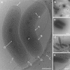
FIG. 1.
FIG. 1.
Intracellular structure and variation in the shape of B. bacteriovorus. (a) Low-dose, energy-filtered, projection EM images of vitrified cells allow detection of key intracellular components, as indicated by arrows. A, outer membrane; B, inner membrane; C, sheathed flagellum; D, rotor complex; E, chemotaxis receptor array; F, dense granule; G, nucleoid; H, macromolecular complexes; I, peptidoglycan layer; J, needlelike structures at the anterior pole. (a to e) Examples illustrating the flexibility of B. bacteriovorus cells, which range from cells with moderate bends (a) to U-shaped bends (b) to cells distorted by virtue of intercellular contact (c) and cell edges flattened by contact with the carbon substrate. The large black dots at the top and bottom right of the images are 15-nm gold particles deposited on the carbon film and used as fiducial markers to align multiple images in a tilt series. (a and c to e) Scale bar = 200 nm. (b) Scale bar = 1 μm.
FIG. 2.
FIG. 2.
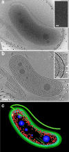
Cellular interior revealed by cryo-electron tomography. (a) Projection image recorded as part of a tilt series for a specimen of plunge-frozen B. bacteriovorus cells. (b) Central 8-nm slice through the reconstructed 3D volume obtained from the same tilt series. (c) 3D rendering of key cellular structures segmented from the tomographic volume. The flagellum, inner and outer membranes, and filamentlike structures in the cytoplasm are green, putative ribosomes are red, and dense granules are blue. The inset in panel a shows a projection image with periodic, 26-Å-spaced striations in the nucleoid region, consistent with the expected packing arrangement of DNA. The inset in panel b shows the anterior end of the cell, including structures (indicated by arrowheads) that protrude outward and may be relevant for making contact with prey. (a and b) Scale bars = 150 nm. (Panel a inset) Scale bar = 10 nm. (Panel b inset) Scale bar = 25 nm.
FIG. 3.
FIG. 3.
Elemental analysis of Bdellovibrio dense granules. (a) Projection image of a cell recorded using scanning transmission EM. Circles 1, 2, and 3 indicate the centers of square regions (20 by 20 nm) analyzed by EELS to determine the elemental composition of the dense granules, the surrounding nucleoid, and the extracellular medium, respectively. (b) EELS spectra of the three regions, indicating that the dense granules are enriched in phosphorus, oxygen, and calcium. See Materials and Methods for additional details.
FIG. 4.
FIG. 4.
Distribution of the locations and sizes of the dense granules along the length of the cell. (a) The data show that the dense granules are not present at specific positions relative to the pole of the cell and can be various sizes. (b) Correlation between the size of the dense granules and the size of the cell, expressed in granule size/cell length, indicating that there is no obvious correlation between the size of the cells and the overall size of the granules.
FIG. 5.
FIG. 5.
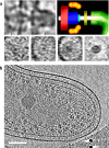
Visualization of the motility and chemotaxis machinery of B. bacteriovorus. (a) (Top left panel) Eight-nanometer slice extracted from a tomographic volume in an orientation coplanar with the axis of the rotor complex. Sections of the individual elements of the ring are visible. (Top right panel) Schematic diagram of the tomogram corresponding to the slice shown in the top left panel showing components expected to be found in a single rotor complex. (Bottom panels) Four orthogonal slices across the rotor complex shown in the top left panel at places indicated by the tick marks. The density resulting from the flagellum is visible as a small dark ring in the fourth bottom panel and is white in the schematic diagram in the top right panel; a faintly visible density corresponding to the flagellar sheath is green in the top right panel. The density from the flagellum is also visible at the center of the third bottom panel along with contributions from the density indicated by yellow in the top right panel and likely results from the P-ring. The much darker density in the outermost regions of the third panel is due to the outer membrane, and the level is roughly the same as the level of density indicated by yellow in the top right panel. The densities in the first and second panels indicated by red, blue, and orange in the schematic diagram likely correspond to contributions from the C-ring, the MS-ring, and the MotAB complex, respectively. (b) Eight-nanometer tomographic slice through the center of the same cell near the flagellar pole, showing the spatial arrangement of the chemotaxis receptor array (visible as a band in the cytoplasm [dark line of density inside the inner membrane]) relative to the pole. Scale bar = 50 nm.
FIG. 6.
FIG. 6.
Cryo-electron tomography of vitrified B. bacteriovorus cells shaped around the edges of the carbon film-vitreous ice interface. Panels a to d and panels e to h show two examples. (a and e) Low-dose projection images; (b, c, f, and g) 6-nm-thick tomographic slices from different depths of the cellular tomograms; (d and h) segmented rendering of portions of the bent cells, showing the wrapping of the cells around the edge of the carbon film. Scale bars = 200 nm. The inset in panel c shows that despite extensive changes in curvature, the spacing between the inner and outer membranes is maintained along the length of the bacterium.
FIG. 7.
FIG. 7.
Visualization of cytoskeletal elements in the cytoplasm of B. bacteriovorus: tomographic slices showing the presence of bundles of filaments oriented parallel (a and b) and transverse (indicated by the circle) (c) to the plane of the plasma membrane. Scale bars = 100 nm.

References

    1. Abram, D., and B. K. Davis. 1970. Structural properties and features of parasitic Bdellovibrio bacteriovorus. J. Bacteriol. 104948-965. - PMC - PubMed
    1. Abram, D., J. Castro e Melo, and D. Chou. 1974. Penetration of Bdellovibrio bacteriovorus into host cells. J. Bacteriol. 118663-680. - PMC - PubMed
    1. Bobyk, M. A., A. V. Afinogenova, M. V. Dudinskaya, V. A. Lambina, and I. S. Kulaev. 1980. Detection of polyphosphates and enzymes of polyphosphate metabolism in Bdellovibrio bacteriovorus. Zentralbl. Bakteriol. Naturwiss. 135461-466. - PubMed
    1. Briegel, A., D. P. Dias, Z. Li, R. B. Jensen, A. S. Frangakis, and G. J. Jensen. 2006. Multiple large filament bundles observed in Caulobacter crescentus by electron cryotomography. Mol. Microbiol. 625-14. - PubMed
    1. Brown, M. R., and A. Kornberg. 2004. Inorganic polyphosphate in the origin and survival of species. Proc. Natl. Acad. Sci. USA 10116085-16087. - PMC - PubMed

Publication types

MeSH terms

LinkOut - more resources