Review
. 2008 Feb;9(2):129-36.
doi: 10.1038/ni1560.

Of lineage and legacy: the development of mammalian hematopoietic stem cells

Affiliations
Review

Of lineage and legacy: the development of mammalian hematopoietic stem cells

Elaine Dzierzak et al. Nat Immunol. 2008 Feb.

Abstract

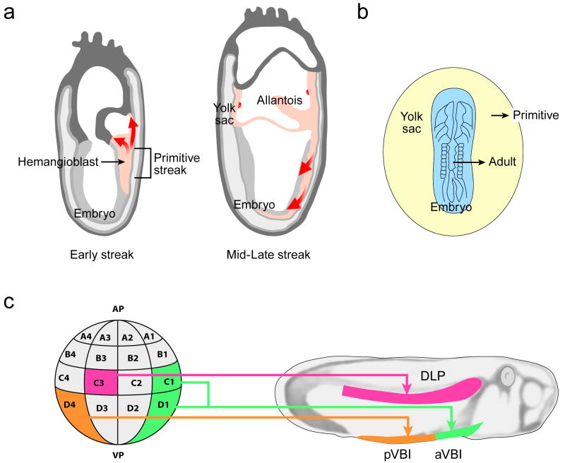
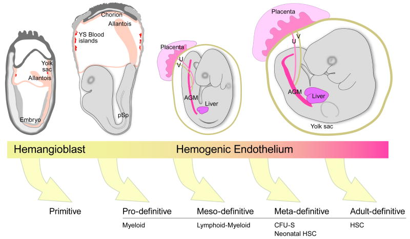
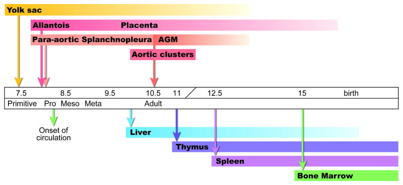
The hematopoietic system is one of the first complex tissues to develop in the mammalian conceptus. Of particular interest in the field of developmental hematopoiesis is the origin of adult bone marrow hematopoietic stem cells. Tracing their origin is complicated because blood is a mobile tissue and because hematopoietic cells emerge from many embryonic sites. The origin of the adult mammalian blood system remains a topic of lively discussion and intense research. Interest is also focused on developmental signals that induce the adult hematopoietic stem cell program, as these may prove useful for generating and expanding these clinically important cell populations ex vivo. This review presents a historical overview of and the most recent data on the developmental origins of hematopoiesis.


Figures

Figure 1
Figure 1
Vertebrate hematopoietic development. (a) Mesodermal migration during the early streak (left panel) and mid/late streak stage (right panel) in the mouse conceptus. In the early streak stage, mesoderm emerging from the primitive streak forms the extraembryonic yolk sac and slightly later, the allantois. At the mid/late streak stage, mesoderm emerging from the anterior primitive streak forms the axial, paraxial and lateral mesoderm in the rostral region of the embryo. The mesoderm from the posterior primitive streak forms the paraxial and lateral mesoderm of the remaining trunk region of the embryo. The red arrows depict the emigration of mesoderm after egressing from the primitive streak. (b) Inter- and intra-species grafting in early pre-circulation avian embryos (for example a quail embryo body grafted onto a chick yolk sac) revealed the origins of the adult blood in the embryo body and not the yolk sac. (c) Amphibian embryos at the 32 cell stage (left) were genetically marked and the progeny traced to larval stages (right). The C3 blastomere gives rise to the DLP (dorsal lateral plate) mesoderm and more specifically to the dorsal aorta and the hematopoietic clusters within the lumen. The D4 blastomere gives rise to the pVBI (posterior ventral blood island) and the C1 + D1 blastomeres to the aVBI (anterior ventral blood island). Drawings adapted from , ; and the Edinburgh mouse atlas website (http://genex.hgu.mrc.ac.uk/).
Figure 2
Figure 2
Mouse conceptuses at E (embryonic day) 7.5, 8.25, 9.0 and 10.5 (respectively from left to right). At least five classes of hematopoietic cells (as defined by function) are progressively generated in the mouse conceptus as indicated by the arrows. The primitive class is derived from hemangioblasts and the pro, meso, meta and adult classes are thought to be derived from specialized vascular cells called hemogenic endothelium. Some of these cells are derived from distinct mesodermal lineages emigrating from the primitive streak (see Fig. 1). The E7.5 and E8.25 conceptuses show the outgrowing allantois that will fuse with the chorion to form the placenta. The circulation is established at E8.25-8.5. The E9 embryo has turned and is enveloped in the yolk sac. Colonization of the liver by hematopoietic progenitors begins at late E9. The E10.5 conceptus contains hematopoietic clusters in the dorsal aorta within the AGM regions, the vitelline (V) and umbilical (U) arteries and the first adult HSCs are found in these vessels. Drawings adapted from the Edinburgh mouse atlas website (http://genex.hgu.mrc.ac.uk/).
Figure 3
Figure 3
Time line of hematopoietic events in the mouse conceptus. The arrows above the time line indicate the onset of specific hematopoietic cell generation and/or appearance and the arrows below indicate the earliest time of colonization of the secondary hematopoietic territories.

References

    1. Sabin F. Studies on the origin of blood vessels and of red blood corpuscles as seen in the living blastoderm of chicks during the second day of incubation. Carnegie Inst Wash Pub # 272, Contrib Embryol. 1920;9:214.
    1. Murray P. The development in vitro of the blood of the early chick embryo. Proc Roy Soc London. 1932;11:497–521.
    1. Choi K, Kennedy M, Kazarov A, Papadimitriou JC, Keller G. A common precursor for hematopoietic and endothelial cells. Development. 1998;125:725–732. - PubMed
    1. Fehling HJ, et al. Tracking mesoderm induction and its specification to the hemangioblast during embryonic stem cell differentiation. Development. 2003;130:4217–4227. - PubMed
    1. Huber TL, Kouskoff V, Fehling HJ, Palis J, Keller G. Haemangioblast commitment is initiated in the primitive streak of the mouse embryo. Nature. 2004;432:625–630. - PubMed

Publication types