Review
. 2008 Mar;153 Suppl 1(Suppl 1):S137-53.
doi: 10.1038/sj.bjp.0707659. Epub 2008 Jan 21.

Glycogen synthase kinase 3 (GSK3) in the heart: a point of integration in hypertrophic signalling and a therapeutic target? A critical analysis

Affiliations
Review

Glycogen synthase kinase 3 (GSK3) in the heart: a point of integration in hypertrophic signalling and a therapeutic target? A critical analysis

P H Sugden et al. Br J Pharmacol. 2008 Mar.

Abstract

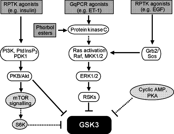
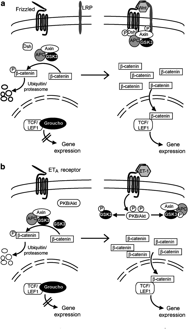
Glycogen synthase kinase 3 (GSK3, of which there are two isoforms, GSK3alpha and GSK3beta) was originally characterized in the context of regulation of glycogen metabolism, though it is now known to regulate many other cellular processes. Phosphorylation of GSK3alpha(Ser21) and GSK3beta(Ser9) inhibits their activity. In the heart, emphasis has been placed particularly on GSK3beta, rather than GSK3alpha. Importantly, catalytically-active GSK3 generally restrains gene expression and, in the heart, catalytically-active GSK3 has been implicated in anti-hypertrophic signalling. Inhibition of GSK3 results in changes in the activities of transcription and translation factors in the heart and promotes hypertrophic responses, and it is generally assumed that signal transduction from hypertrophic stimuli to GSK3 passes primarily through protein kinase B/Akt (PKB/Akt). However, recent data suggest that the situation is far more complex. We review evidence pertaining to the role of GSK3 in the myocardium and discuss effects of genetic manipulation of GSK3 activity in vivo. We also discuss the signalling pathways potentially regulating GSK3 activity and propose that, depending on the stimulus, phosphorylation of GSK3 is independent of PKB/Akt. Potential GSK3 substrates studied in relation to myocardial hypertrophy include nuclear factors of activated T cells, beta-catenin, GATA4, myocardin, CREB, and eukaryotic initiation factor 2Bvarepsilon. These and other transcription factor substrates putatively important in the heart are considered. We discuss whether cardiac pathologies could be treated by therapeutic intervention at the GSK3 level but conclude that any intervention would be premature without greater understanding of the precise role of GSK3 in cardiac processes.


Figures

Figure 1
Figure 1
Regulation of glycogen metabolism by glycogen synthase kinase 3 (GSK3). (a) Casein kinase 2 (CK2) phosphorylates a priming Ser residue in the C terminus of glycogen synthase (Ser657 in the muscle isoform of Homo sapiens) to initiate a relay of GSK3-catalysed phosphorylations, as described in the text. (b) Glycogen synthase activity is regulated by a phosphorylation–dephosphorylation cycle. In the preprandial state, plasma insulin concentrations are low and GSK3 is active. GSK3 phosphorylates glycogen synthase and converts it to the less-active (glucose-6-phosphate (G6P) dependent) form, thus inhibiting glycogen synthesis. When plasma insulin concentrations rise after feeding, insulin promotes phosphorylation of GSK3 and inhibits GSK3 activity. Dephosphorylation of glycogen synthase by protein phosphatases (GS phosphatase) subsequently increases the activity of glycogen synthase and promotes glycogen synthesis. (c) GSK3α and GSK3β exhibit a high degree of homology in their kinase domains, diverging at their N- and C-terminal regions. GSK3α contains an N-terminal Gly-rich domain of unknown function. However, Gly-rich domains are classically associated with binding of nucleotides (Bossemeyer, 1994), though this domain is distinct from the ATP-binding site in the kinase domain. Both isoforms are constitutively phosphorylated on a Tyr residue in the kinase domain and this phosphorylation is required for activity. Phosphorylation of a conserved N-terminal Ser residue inhibits the activity of GSK3.
Figure 2
Figure 2
Signalling pathways leading to the phosphorylation and inhibition of glycogen synthase kinase 3 (GSK3). The following scheme is a simplification of the events and only the salient features of the pathways are outlined. Receptor protein tyrosine kinase (RPTK) agonists (for example, insulin) stimulate the activity of phosphoinositide 3′ kinase (PI3K), which phosphorylates the membrane phospholipid phosphatidylinositol 4,5-bisphosphate (PtdIns(4,5)P2), to produce phosphatidylinositol 3,4,5-trisphosphate (PtdInsP3). PtdInsP3 remains in the plane of the membrane, activating 3-phosphoinositide-dependent kinase 1 (PDK1) and also causing translocation of protein kinase B/Akt (PKB/Akt) to the membrane where it is phosphorylated on Thr308 and partially activated by PDK1. Phosphorylation of PKB/Akt(Ser473) by the mammalian target-of-rapamycin complex 2 (not shown) fully activates the kinase. PKB/Akt subsequently phosphorylates the two GSK3 isoforms on a Ser-residue in each of their N-terminal domains to inhibit their activities (see Figure 1c). Gq-protein-coupled receptor (GqPCR) agonists (for example, endothelin-1 (ET-1)) stimulate phospholipase Cβ which hydrolyses PtdIns(4,5)P2 to produce diacylglycerol and inositol 1,4,5-trisphosphate. The former is the physiological activator of the diacylglycerol-regulated protein kinase C isoforms (which can also be activated by the tumour-promoting phorbol esters), whereas the latter is important in the regulation of intracellular Ca2+ movements. Protein kinase C activates the extracellular signal-regulated kinases 1/2 (ERK1/2) cascade, which involves the small guanine nucleotide-binding protein Ras. Conversion of Ras.GDP to its biologically active form, Ras.GTP, leads to the hierarchical activation of three levels of protein kinases. Raf phosphorylates and activates mitogen-activated protein kinase kinases 1/2 (MKK1/2), which then phosphorylate and activate ERK1/2. The signalling pathway then diverges. One consequence of activation of ERK1/2 is their phosphorylation and activation of the p90-ribosomal subunit S6 kinases (RSKs), which phosphorylate and inhibit GSK3. Some RPTK agonists (for example, epidermal growth factor, EGF), in addition to activating PKB/Akt (Clerk et al., 2006), activate the ERK1/2 cascade by stimulating GTP/GDP exchange on Ras via the Grb2/Sos guanine nucleotide exchange factor system, and thus activate Ras. In addition, PKB/Akt also activates the mammalian target-of-rapamycin complex 1 (mTOR). This leads to phosphorylation and activation of p70/p85-ribosomal subunit S6 kinases (S6Ks), thence to phosphorylation and inhibition of GSK3. Agents that raise cyclic AMP concentrations and activate cyclic AMP-dependent protein kinase A (PKA) may bring about phosphorylation and inhibition of GSK3, though the physiological significance of this mode of regulation is somewhat obscure.
Figure 3
Figure 3
Regulation of the transcriptional co-regulator β-catenin. (a) Glycogen synthase kinase 3 (GSK3) participates in the regulation of the canonical Wnt pathway. In the absence of Wnt, a pool of GSK3 is present in a complex with axin and the adenomatous polyposis coli (APC) protein. In this complex, GSK3 is catalytically active and it phosphorylates previously ‘primed' β-catenin (that is, phosphorylated by casein kinase 1 on Ser45) at three further sites: Thr41, Ser37 and Ser33. This polyphospho-β-catenin is then degraded through the ubiquitin–proteasome system. When Wnts engage to the extracellular domain of their transmembrane receptors (Frizzled) and to the lipoprotein receptor-related protein (LRP) co-receptor, the axin–APC–GSK3 complex in association with the scaffold protein Dishevelled (Dsh) binds to the intracellular domains of Frizzled and LRP, and phosphorylations occur. GSK3 is now unable to phosphorylate β-catenin, which accumulates and enters the nucleus. Here, it displaces the co-repressor Groucho from its complex with T-cell-specific transcription factor/lymphoid enhancer binding factor 1 (TCF/LEF1), initiating gene expression. (b) It is suggested (Shevtsov et al., 2006) that a similar scheme may operate when endothelin-1 (ET-1) binds to its receptor in cardiac myocytes (the ETA receptor seems to be the principal endothelin-1 receptor). This leads to phosphorylation and activation of protein kinase B/Akt (PKB/Akt) (Figure 2) which then phosphorylates and inhibits GSK3 promoting β-catenin accumulation. It is however unclear which pool(s) of GSK3 is/are involved in this particular phosphorylation of β-catenin.
Figure 4
Figure 4
Potential glycogen synthase kinase (GSK3) Ser/Thr-directed phosphorylation relays in nuclear factor of activated T cells (NFATs). (a) Sequences for Homo sapiens NFATs were aligned by CLUSTAL W (1.83). Accession numbers are NFATc1, NP_006153.2 (825 residues); NFATc2, NP_036472.2 (921 residues); NFATc3, NP_004546.1 (1068 residues) and NFATc4, NP_004545.2 (902 residues). Alignment of the ‘atypical' NFAT, NFAT5, has been omitted. The extended and highly conserved Ser/Thr relays (potentially phosphorylated by GSK3) present in the N-terminal regions of NFATc1, NFATc2, NFATc3 and NFATc4 are in underlined, bold italicized type. In NFATc1, these sites are thought to be phosphorylated by GSK3. Other more limited relays are present in individual NFATs and all contain a Ser-rich domain N-terminal to the GSK3 consensus sequences that is phosphorylated by casein kinase 1. (b) GSK3 relays (underlined, bold italicized type) present in GATA4(1–217). (c) GSK3 relays (underlined, bold italicized type) in mouse myocardin (residues 454–466 and 624–636) as identified by Badorff et al. (2005). Identical sequences are present in H. sapiens myocardin. There are also ‘out-of-phase' relays in these regions (shaded, bold and italicized), which are not as strongly conserved in H. sapiens myocardin.
Figure 5
Figure 5
Relevant aspects of phosphoinositide metabolism. Phosphatidylinositol 4,5-bisphosphate (PtdIns(4,5)P2) is an important phospholipid in cell signalling producing three separate ‘second messenger' molecules. It is hydrolysed by a variety of phospholipase C (PLC) isoforms to produce diacylglycerol and inositol 1,4,5-trisphosphate. The former is the physiological activator of the diacylglycerol-regulated protein kinase C isoforms (see Figure 2), whereas the latter is important in the regulation of intracellular Ca2+ movements. In cardiac myocytes, the phospholipase Cβ isoforms are best studied in this regard. In a second signalling pathway, PtdIns(4,5)P2 is phosphorylated by phosphoinositide 3′ kinase (PI3K) to form phosphatidylinositol 3,4,5-trisphosphate (PtdIns(3,4,5)P3). This regulates the activity of the 3-phosphoinositide-dependent kinase/protein kinase B (Akt) signalling pathway (see Figure 2). Phosphoinositides are dephosphorylated (hydrolysed) by a number of lipid phosphatases. One such phosphatase which hydrolyses the 5-phosphate of phosphatidylinositol 4,5-bisphosphate and PtdIns(3,4,5)P3 is inositol polyphosphate-5-phosphatase f, which produces phosphatidylinositol 4-phosphate and phosphatidylinositol 3,4-bisphosphate, respectively.

References

    1. Adams JW, Sakata Y, Davis MG, Sah VP, Wang Y, Liggett SB, et al. Enhanced Gαq signaling: a common pathway mediates cardiac hypertrophy and apoptotic heart failure. Proc Natl Acad Sci USA. 1998;95:10140–10145. - PMC - PubMed
    1. Akhter SA, Luttrell LM, Rockman HA, Iaccarino G, Lefkowitz RJ, Koch WJ. Targeting the receptor–Gq interface to inhibit in vivo pressure overload myocardial hypertrophy. Science. 1998;280:574–577. - PubMed
    1. Ali A, Hoeflich KP, Woodgett JR. Glycogen synthase kinase-3: properties, functions, and regulation. Chem Rev. 2001;101:2527–2540. - PubMed
    1. Allen DL, Harrison BC, Maass A, Bell ML, Byrnes WC, Leinwand LA. Cardiac and skeletal muscle adaptations to voluntary wheel running in the mouse. J Appl Physiol. 2001;90:1900–1908. - PubMed
    1. Andersen JB, Rourke BC, Caiozzo VJ, Bennett AF, Hicks JW. Postprandial hypertrophy in pythons. Nature. 2005;434:37–38. - PubMed

MeSH terms