. 2008 Mar;36(3):415-24.
doi: 10.1007/s10439-007-9385-8. Epub 2008 Jan 18.

Biomechanical analysis of reducing sacroiliac joint shear load by optimization of pelvic muscle and ligament forces

Affiliations

Biomechanical analysis of reducing sacroiliac joint shear load by optimization of pelvic muscle and ligament forces

J J M Pel et al. Ann Biomed Eng. 2008 Mar.

Abstract

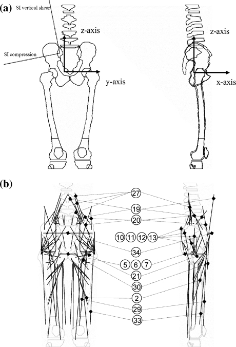
Effective stabilization of the sacroiliac joints (SIJ) is essential, since spinal loading is transferred via the SIJ to the coxal bones, and further to the legs. We performed a biomechanical analysis of SIJ stability in terms of reduced SIJ shear force in standing posture using a validated static 3-D simulation model. This model contained 100 muscle elements, 8 ligaments, and 8 joints in trunk, pelvis, and upper legs. Initially, the model was set up to minimize the maximum muscle stress. In this situation, the trunk load was mainly balanced between the coxal bones by vertical SIJ shear force. An imposed reduction of the vertical SIJ shear by 20% resulted in 70% increase of SIJ compression force due to activation of hip flexors and counteracting hip extensors. Another 20% reduction of the vertical SIJ shear force resulted in further increase of SIJ compression force by 400%, due to activation of the transversely oriented M. transversus abdominis and pelvic floor muscles. The M. transversus abdominis crosses the SIJ and clamps the sacrum between the coxal bones. Moreover, the pelvic floor muscles oppose lateral movement of the coxal bones, which stabilizes the position of the sacrum between the coxal bones (the pelvic arc). Our results suggest that training of the M. transversus abdominis and the pelvic floor muscles could help to relieve SI-joint related pelvic pain.


Figures

Figure 1
Figure 1
Panel (a) shows the bones on which the muscles, ligaments and joint reaction forces act in the frontal plane (left) and the median plane (right): the lowest thoracic vertebra, five lumbar vertebrae, the sacrum, the left and right coxal bones, and the left and right femurs. The coordinate system is defined with the origin halfway between the rotation centers of the hip joints. Axes: x posterior, y left, z vertical. Panel (b) shows, superimposed on the bones, the vectors representing the most important force components in the frontal plane (left) and the median plane (right), see also Table 1. The labels in this panel refer to a selection of the muscle structures listed in Table 1
Figure 2
Figure 2
Directions of the force in the frontal plane exerted by the right ilium through the SIJ on the sacrum as a reaction to trunk load, Ftrunk. Panel (a): initial loading condition without limitation of the vertical shear component (563 N, see under “initial” in Table 2). This condition led to loading of the sacrotuberal ligaments, Fsacrotuberal lig. (solid thick line). Panel (b): loading condition with the vertical shear component preset at a 120 N lower level than the initial value (see under 443 N in Table 2). This condition led to loading of the sacrospinal ligaments, Fsacrospinal lig. (solid thick line). Panel (c): loading condition with the vertical shear component preset at a 240 N lower level than the initial value (see under 323 N in Table 2). In this situation, SIJ compression force increased by ∼400%, mainly by M. transversus abdominis, Ftransversus abdominis, and the pelvic floor, Fpelvic floor, muscle forces. The location of these muscles is schematically drawn by the thick solid lines, including the M. pubococcygeus, the M. iliococcygeus and the M. coccygeus (as drawn from the mid to the lateral position). It also led to loading of the iliolumbar ligaments to the maximum allowed force Filiolumbar lig. of 250 N, (solid thick line). 3-D images copyright of Primal Pictures Ltd. http://www.primalpictures.com
Figure 3
Figure 3
Analogy of pelvic bones supporting the trunk with a classical stone arc. The M. transversus abdominis and the pelvic floor muscles caudal to the SIJ mainly oppose lateral movement of the coxal bones. Spinal loading is transferred mainly by compression forces through the SIJ to the coxal bones and further down to the legs. 3-D images copyright of Primal Pictures Ltd. http://www.primalpictures.com
Figure 4
Figure 4
A flow chart of the optimization scheme used to calculate the optimum set of muscle, ligament and joint reaction forces on the basis of geometry, positions, external forces, and force limitations

References

    1. {'text': '', 'ref_index': 1, 'ids': [{'type': 'DOI', 'value': '10.1016/0268-0033(95)90432-9', 'is_inner': False, 'url': 'https://doi.org/10.1016/0268-0033(95)90432-9'}, {'type': 'PubMed', 'value': '11415526', 'is_inner': True, 'url': 'https://pubmed.ncbi.nlm.nih.gov/11415526/'}]}
    2. Adams M. A., Dolan P. Recent advances in lumbar spinal mechanics and their clinical significance Clin. Biomech. 10:3–19, 1995 - PubMed
    1. {'text': '', 'ref_index': 1, 'ids': [{'type': 'DOI', 'value': '10.1016/0021-9290(85)90012-0', 'is_inner': False, 'url': 'https://doi.org/10.1016/0021-9290(85)90012-0'}, {'type': 'PubMed', 'value': '4055812', 'is_inner': True, 'url': 'https://pubmed.ncbi.nlm.nih.gov/4055812/'}]}
    2. Anderson C. K., Chaffin D. B., Herrin G. D., Matthews L. S. A biomechanical model of the lumbosacral joint during lifting activities. J. Biomech. 18:571–584, 1985 - PubMed
    1. {'text': '', 'ref_index': 1, 'ids': [{'type': 'DOI', 'value': '10.1016/0268-0033(96)00033-2', 'is_inner': False, 'url': 'https://doi.org/10.1016/0268-0033(96)00033-2'}, {'type': 'PubMed', 'value': '11415651', 'is_inner': True, 'url': 'https://pubmed.ncbi.nlm.nih.gov/11415651/'}]}
    2. Andersson E. A., Oddsson L. I. E., Grundstrom H., Nilsson J., Thorstensson A. EMG activities of the quadratus lumborum and erector spinae muscles during flexion–relaxation and other motor tasks. Clin. Biomech. 11:392–400, 1996 - PubMed
    1. {'text': '', 'ref_index': 1, 'ids': [{'type': 'PubMed', 'value': '7154650', 'is_inner': True, 'url': 'https://pubmed.ncbi.nlm.nih.gov/7154650/'}]}
    2. Brand R. A., Crowninshield R. D., Wittstock C. E., Pedersen D. R., Clark C. R., van Krieken F. M. A model of lower extremity muscular anatomy J. Biomech. 104:304–310, 1982 - PubMed
    1. {'text': '', 'ref_index': 1, 'ids': [{'type': 'DOI', 'value': '10.1016/0021-9290(86)90164-8', 'is_inner': False, 'url': 'https://doi.org/10.1016/0021-9290(86)90164-8'}, {'type': 'PubMed', 'value': '3771581', 'is_inner': True, 'url': 'https://pubmed.ncbi.nlm.nih.gov/3771581/'}]}
    2. Brand R. A., Pedersen D. R., Friederich J. A. The sensitivity of muscle force predictions to changes in physiologic cross-sectional area. J. Biomech. 19:589–596, 1986 - PubMed