. 2008 Jul;124(3):322-8.
doi: 10.1111/j.1365-2567.2007.02789.x. Epub 2008 Jan 18.

Conformation of human leucocyte antigen-C molecules at the surface of human trophoblast cells

Affiliations

Conformation of human leucocyte antigen-C molecules at the surface of human trophoblast cells

Richard Apps et al. Immunology. 2008 Jul.

Abstract

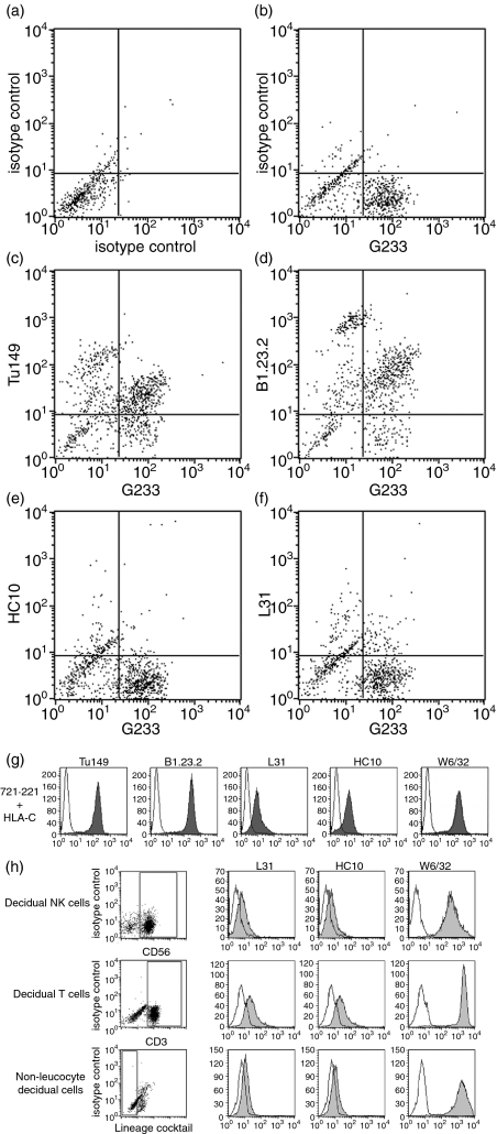
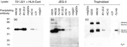
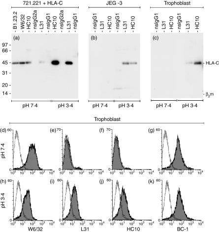
Human leucocyte antigen (HLA)-C is expressed at lower levels than other classical HLA-I molecules on somatic cells. Surface HLA-C proteins can occur as conventionally beta(2)-microglobulin (beta2m)-associated complexes or as open conformers dissociated from peptide and/or beta(2)m. We investigated the conformation of HLA-C molecules on normal human trophoblast cells, which invade the maternal decidua during placentation. A panel of monoclonal antibodies to different conformations of HLA-I molecules was used in flow cytometry and surface immunoprecipitation experiments. On the surface of trophoblast cells only beta(2)m-associated complexes of HLA-C molecules were detected. In contrast, both open conformers and beta(2)m-associated HLA-C could be detected on other cells from the decidua, HLA-C-transfectants and cell lines. The levels of HLA-C expressed on primary trophoblast cells could be detected by antibodies specific to non-beta(2)m-associated conformations because binding was seen after acid-induced denaturation of surface proteins. In contrast to HLA-G molecules on trophoblasts, we found no evidence for the presence of disulphide-linked multimers of HLA-C complexes. These results show that most HLA-C molecules present at the trophoblast cell surface are in the conventional beta(2)m-associated conformation. These findings have implications regarding the stability of trophoblast HLA-C molecules and how they interact with receptors on decidual leucocytes during placentation.


Figures

Figure 1
Figure 1
Surface human leucocyte antigen class I (HLA-I) molecules on primary cells from normal human pregnancies. Compared to isotype controls (a), extravillous trophoblast cells are identified by staining with the HLA-G specific monoclonal antibody (mAb) G233 (b). Double labelling with the mAb Tü149 (c) or B1.23.2 (d) confirms that these HLA-G+ cells also express HLA-C in the β2microglobulin-associated conformation. mAb HC10 (e) or L31 (f) that bind open conformers of HLA-C molecules do not bind trophoblast. The mAb recognizing all conformations of HLA-C molecules detect their antigens at the surface of 721.221 cells transfected with HLA-Cw4 (g). The mAb HC10 and L31 also detect their antigens on primary decidual cells identified by gates for CD56+, CD3+ or cells negative for leucocyte lineage markers (h). In histograms binding of the indicated mAb is shown in grey compared to staining of an isotype control in white.
Figure 2
Figure 2
Immunoprecipitation and Western blotting of surface-biotinylated human leucocyte antigen class I (HLA-I) molecules on trophoblast cells. 721.221 cells transfected with HLA-Cw4 (a), the JEG-3 choriocarcinoma line (b) and human trophoblast cells (c) were surface biotinylated, lysed and immunoprecipitated with the indicated monoclonal antibody (mAb). Precipitated complexes were resolved by non-reducing sodium dodecyl sulphate–polyacrylamide gel electrophoresis and surface complexes were detected by streptavidin-horseradish peroxidase. (a) From transfectants 45 000 Da molecular weight HLA-C molecules are detected by mAb to both β2microglobulin-associated (W6/32, B1.23.2, Tü149) as well as open conformations (L31, HC10) of the HLA-C molecule. (b,c) From both JEG-3 and primary trophoblast cells, W6/32, B1.23.2 and Tü149 mAb precipitate the 45 000 Da molecular weight HLA-C molecule. W6/32 and G233 also detect the 39 000 Da molecular weight HLA-G molecule expressed by trophoblast and JEG-3 cells. L31 and HC10 mAb do not detect their antigen on trophoblast cells and only very weakly on JEG-3 cells. Gels in this figure are each representative of three independent experiments, using in total trophoblast pooled from nine individual samples.
Figure 3
Figure 3
Monoclonal antibody (mAb) to open conformers of human leucocyte antigen (HLA)-C bind their antigen on trophoblast cells after acid-induced denaturation of the HLA-I complexes. The mAb binding after a brief acid incubation was investigated by immunoprecipitation (a–c) and flow cytometry (d–k). (a) For HLA-C transfected cells, mAb recognizing both β2microgobulin-associated (W6/32, B1.23.2) and open conformations (L31, HC10) of HLA-C immunoprecipitate their 45 000 Da molecular weight antigen from untreated lysates. The mAb to open conformers clearly detect more antigen after acid treatment. (b,c) L31 and HC10 only immunoprecipitate their 45 000 Da molecular weight antigen from acidified lysates of JEG-3 (b) and primary trophoblast cells (c), but not from untreated lysates. Flow cytometry staining of HLA-G+ cells gated from the primary trophoblast preparations is shown, for cells kept at pH 7·4 (d–g) or briefly incubated in acidic conditions (h–k). (d,h) W6/32 mAb staining decreases after acid exposure, consistent with the partial denaturation of some HLA-I complexes. (e, f, i, j) HC10 and L31 mAb clearly bind trophoblast only after acid treatment. (g, k) Detection of the non-HLA-I antigen BC-1 is not influenced by acid treatment.

References

    1. Loke YW, King A, Burrows T, et al. Evaluation of trophoblast HLA-G antigen with a specific monoclonal antibody. Tissue Antigens. 1997;50:135–46. - PubMed
    1. King A, Boocock C, Sharkey AM, Gardner L, Beretta A, Siccardi AG, Loke YW. Evidence for the expression of HLA-C Class I mRNA and protein by human first trimester trophoblast. J Immunol. 1996;156:2068–76. - PubMed
    1. King A, Burrows TD, Hiby SE, et al. Surface expression of HLA-C antigen by human extravillous trophoblast. Placenta. 2000;21:376–87. - PubMed
    1. King A, Allan DS, Bowen M, et al. HLA-E is expressed on trophoblast and interacts with CD94/NKG2 receptors on decidual NK cells. Eur J Immunol. 2000;30:1623–31. - PubMed
    1. Verma S, King A, Loke YW. Expression of killer cell inhibitory receptors on human uterine natural killer cells. Eur J Immunol. 1997;27:979–83. - PubMed

Publication types